眼底
眼底(fundus of the eye)是指与晶状体对面的眼球内表面,包括视网膜、视盘、黄斑、中央凹和后极[1]。眼底可通过检眼镜和/或眼底照相做检查。


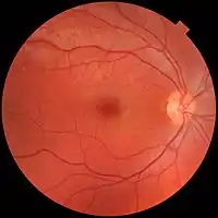
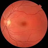
正常眼底的照片,由前方观察受检者右眼(左图)左眼(右图),黄斑(图中暗影)位于影像中央,视盘(图中亮影)则近鼻侧(两图之间)。两视盘外侧(颞侧)边缘均有色素沉着,此非病理现象。右眼(左图)大血管流域附近有较明显的浅色区域,这也是正常现象,常见于年轻人。
临床意义
可通过眼底观察(检眼镜、眼底镜检查)检测到的医学体征包括出血、渗出液、棉絮斑、血管异常(纡曲、搏动和新生血管)和色素沉着。[2]在高血压视网膜病变中,可以看到小动脉缢缩,如 "银线"(silver wiring),以及血管纡曲等现象。
眼底是人体唯一可以直接观察微循环的部位。视盘周围的血管直径约为 150 μm,使用检眼镜可观察直径小至 10 μm 的血管[3]。
參考
- Cassin, B. and Solomon, S. Dictionary of Eye Terminology. Gainesville, Florida: Triad Publishing Company, 1990.
- Baker ML, Hand PJ, Wang JJ, Wong TY. Retinal Signs and Stroke. Stroke. 2008;39(4):1371-1379. doi:10.1161/strokeaha.107.496091
- Ronald Pitts Crick, Peng Tee Khaw, A Textbook of Clinical Ophthalmology: A Practical Guide to Disorders of the Eyes and Their Management, 3rd edition, World Scientific, 2003, ISBN 981-238-128-7
This article is issued from Wikipedia. The text is licensed under Creative Commons - Attribution - Sharealike. Additional terms may apply for the media files.