超声心动图
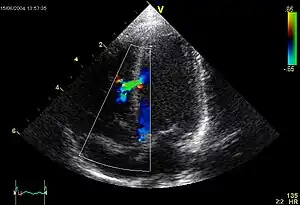
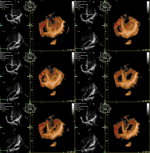
超声心动图,是一种心脏超声波检查,它使用标准的超声波技术或多普勒超声显示心脏的二维图片[1]。现在最新的超声诊断系统采用三维即时成像。耗時大約15-20分鐘,甚至更長。

传统二维超声心动图

三维实时成像
除了产生心血管系统的两维图像,超声心动图使用脉冲或者连续超声波,能对任意位置的血液和心肌组织速度作出准确的测量。这样可以检测心脏瓣膜区域功能、左右侧心脏不正常联系、瓣膜逆流、以及心脏输出量的计算等。其他测量的参数包括心脏尺寸(腔室直径和室间隔厚度)和E/A比值。
参考文献
- Cleve, Jayne; McCulloch, Marti L., Nihoyannopoulos, Petros; Kisslo, Joseph , 编, , Echocardiography (Springer International Publishing), 2018: 33–42, ISBN 9783319716176, doi:10.1007/978-3-319-71617-6_2
外部链接
| 维基共享资源上的相关多媒体资源:超声心动图 |
- Echocardiography (Views, normal values, measurements, free software...) - TECHmED (页面存档备份,存于)
- VIRTUAL TEE – online self-study and teaching resource (页面存档备份,存于)
- VIRTUAL Transthoracic Echocardiography - online self-study and teaching resource (页面存档备份,存于)
- echocardia - online self-study and teaching resource (页面存档备份,存于)
- Echobasics – free online echocardiography tutorial (页面存档备份,存于)
- CT2TEE – transesophageal echocardiography simulator (页面存档备份,存于)
This article is issued from Wikipedia. The text is licensed under Creative Commons - Attribution - Sharealike. Additional terms may apply for the media files.