| AGTPBP1 | |||||||||||||||||||||||||||||||||||||||||||||||||||
|---|---|---|---|---|---|---|---|---|---|---|---|---|---|---|---|---|---|---|---|---|---|---|---|---|---|---|---|---|---|---|---|---|---|---|---|---|---|---|---|---|---|---|---|---|---|---|---|---|---|---|---|
| Identifiers | |||||||||||||||||||||||||||||||||||||||||||||||||||
| Aliases | AGTPBP1, CCP1, NNA1, ATP/GTP binding protein 1, CONDCA, ATP/GTP binding carboxypeptidase 1 | ||||||||||||||||||||||||||||||||||||||||||||||||||
| External IDs | OMIM: 606830 MGI: 2159437 HomoloGene: 9067 GeneCards: AGTPBP1 | ||||||||||||||||||||||||||||||||||||||||||||||||||
| |||||||||||||||||||||||||||||||||||||||||||||||||||
| |||||||||||||||||||||||||||||||||||||||||||||||||||
| |||||||||||||||||||||||||||||||||||||||||||||||||||
| |||||||||||||||||||||||||||||||||||||||||||||||||||
| |||||||||||||||||||||||||||||||||||||||||||||||||||
| Wikidata | |||||||||||||||||||||||||||||||||||||||||||||||||||
| |||||||||||||||||||||||||||||||||||||||||||||||||||
ATP/GTP binding protein 1 is gene that encodes the protein known as cytosolic carboxypeptidase 1 (CCP1), originally named NNA1. Mice with a naturally occurring mutation of the Agtpbp1 gene are known as pcd mice (Purkinje cell degeneration). [5]
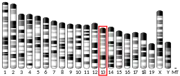
Several spontaneous Agtpbp1 alleles have been discovered in mice.[6] The autosomal recessive Purkinje cell degeneration mutation affects Agtpbp1 located on mouse chromosome 13, with alleles containing a zinc carboxypeptidase domain and an ATP/GTP binding motif, a protein first identified in alpha-motoneurons during axonal regeneration.[7] and destabilized in the mutation.[8]
In Agtpbp1-pcd mutant mice, the predominant pathology comprises near total Purkinje cell loss from the third to the fourth postnatal week along with a more slowly progressing loss in retinal photoreceptors.[9] The slower and less complete degeneration of inferior olive neurons is most probably a consequence of retrograde degeneration secondary to Purkinje cell loss.[10] and the degeneration of deep cerebellar nuclei a consequence of anterograde degeneration secondary to Purkinje cell loss.[11][12]
Based on sequence similarities, CCP1 was proposed to be involved in tubulin processing along with five other carboxypeptidases, designated CCP2 to CCP6.[13] The abnormal development in Purkinje cell dendrites of Agtpbp1-pcd mice was linked to a decrease in microtubule-associated proteins 1B and 2.[14] Microtubule structure and dynamics can be seen to be impaired in embryonic fibroblasts of Agtpbp1-pcd mice.[15] In particular, Purkinje cell loss in Agtpbp1-pcd mice was linked with microtubule hyperglutamylation,[16] as with human subjects lacking Agtpbp1.[17]
In view of Purkinje cell loss, gamma-aminobutyric acid (GABA) concentrations decreased in the cerebellum of Agtpbp1-pcd mutants.[18] In particular, GABA concentrations decreased in the deep cerebellar nuclei, target of Purkinje axons, but not in cerebellar cortex despite Purkinje cell loss, presumably because the maintenance of inhibitory interneurons compensated for it in a shrunken cerebellum.[19] In line with the Purkinje cell loss, the number of GABAergic terminal boutons declined in deep cerebellar nuclei of Agtpbp1-pcd mutants.[20] while the density of GABAergic soma in the deep cerebellar nuclei was normal.[21] Presumably because of post-synaptic supersensitivity following Purkinje cell loss, binding of the GABA-A receptor and its associated benzodiazepine receptor (BZD) receptor increased in deep cerebellar nuclei of Agtpbp1-pcd mutants.[22] In particular, there was an increase in large aggregates of the GABA-A-alpha receptor subtype in deep cerebellar nuclei.[23]
Presumably because of slowly progressing granule cell deterioration.,[24] glutamate concentrations per protein weight decreased in the cerebellar cortex of 6, 9, and 12 month-old Agtpbp1-pcd mutants (McBride and Ghetti, 1978).[25] On the contrary, their glutamate concentrations per tissue weight were equivalent to controls in cerebellar cortex and deep nuclei at younger ages of 1 to 3 months (Roffler-Tarlov et al., 1979),[26] presumably because of slowly progressing granule cell deterioration.[27] Non-NMDA receptor binding decreased in molecular and granule cell layers of the cerebellar cortex but not the deep nuclei of Agtpbp1-pcd mutants (Stasi et al., 1997). More particularly, the decline in binding occurred for the alpha-amino-3-hydroxy-5-methyl-4-isoxazolepropionic acid (AMPA receptor) receptor at the level of molecular and granule cell layers.[28] The residual presence of AMPA receptors in the molecular layer despite Purkinje cell loss was attributed to the continued maintenance of stellate and basket cells.
Monoamine systems have been examined in view of cerebellar targets by 5-hydroxy-tryptamine (5HT) fibers originating from medial and dorsal raphe nuclei, noradrenaline fibers from the locus coeruleus, and dopamine fibers from the ventral tegmental area. 5HT concentrations increased in the cerebellum of Agtpbp1-pcd mutant mice,[29] as did 5HT fiber density.[30] and 5HT uptake binding.[31] The results are more variable when 5HT content per cerebellum is considered, increases being found only in older (9 and 15 months of age) not younger (3 and 6 months of age) mice.[32]
In a similar manner to the 5HT system, noradrenaline concentrations increased in Agtpbp1-pcd mutants.[33][34][35][36] Noradrenaline uptake per protein weight increased in cerebellar cortex and deep nuclei of Agtpbp1-pcd mutants, but was unchanged when surface areas was taken into account.[37] Increases per protein weight were also discerned in the granule cell layer and deep nuclei for alpha-1-adrenergic receptor and alpha-2-adrenergic receptor binding as well as beta-adrenergic receptor binding in cerebellar cortex. With total area binding in cerebellar cortex, values were still higher than normal for alpha-2-adrenergic receptors but were lower than normal for the other two.
More limited data are available with the other major brain catecholamine, dopamine. Dopamine transporter binding increased in deep cerebellar nuclei but decreased in the cerebellar molecular layer of Agtpbp1-pcd mutants.[38]
Agtpbp1-pcd mutant mice display overt ataxia (widespread gait) together with irregularly spaced strides on foot-print analyses[39] and motor coordination deficits based on kinematic analyses of multijoint, interlimb, and whole-body movements,[40] more missteps, shorter steps, and longer step times in the Erasmus ladder task,[41] and latencies before falling from the rotarod performance test and other apparatus.[42][43] The mutants also exhibit anomalies in exploratory activities, including spontaneous alternation.[44][45][46] In addition, Agtpbp1-pcd mice had slowed acquisition of spatial learning in the Morris water maze while swimming normally towards a visible platform relative to heterozygotes but not wild-type mice.[47][48]
Function
CCP1/NNA1 is a zinc carboxypeptidase that contains nuclear localization signals that was initially cloned from regenerating spinal cord neurons of the mouse. Although originally thought to contain an ATP/GTP-binding motif, this was not experimentally verified, and the potential domain is not conserved through evolution.
[supplied by OMIM, Jul 2002].
Notes
References
- 1 2 3 GRCh38: Ensembl release 89: ENSG00000135049 - Ensembl, May 2017
- 1 2 3 GRCm38: Ensembl release 89: ENSMUSG00000021557 - Ensembl, May 2017
- ↑ "Human PubMed Reference:". National Center for Biotechnology Information, U.S. National Library of Medicine.
- ↑ "Mouse PubMed Reference:". National Center for Biotechnology Information, U.S. National Library of Medicine.
- ↑ "Entrez Gene: ATP/GTP binding protein 1". Retrieved 2018-06-13.
- ↑ Fernandez-Gonzalez A, La Spada AR, Treadaway J, Higdon JC, Harris BS, Sidman RL, Morgan JI, Zuo J (2002). "Purkinje cell degeneration (pcd) phenotypes caused by mutations in the axotomy-induced gene, Nna1". Science. 295 (5561): 1904–6. Bibcode:2002Sci...295.1904F. doi:10.1126/science.1068912. PMID 11884758. S2CID 24520602.
- ↑ Harris A, Morgan JI, Pecot M, Soumare A, Osborne A, Soares HD (2000). "Regenerating motor neurons express Nna1, a novel ATP/GTP-binding protein related to zinc carboxypeptidases". Mol Cell Neurosci. 16 (5): 578–96. doi:10.1006/mcne.2000.0900. PMID 11083920. S2CID 32298322.
- ↑ Chakrabarti L, Neal JT, Miles M, Martinez RA, Smith AC, Sopher BL, La Spada AR (2006). "The Purkinje cell degeneration 5J mutation is a single amino acid insertion that destabilizes Nna1 protein". Mamm Genome. 17 (2): 103–110. doi:10.1007/s00335-005-0096-x. PMID 16465590. S2CID 19289988.
- ↑ Mullen RJ, Eicher EM, Sidman RL (1976). "Purkinje cell degeneration: a new neurological mutation in the mouse". Proc Natl Acad Sci USA. 73 (1): 208–12. Bibcode:1976PNAS...73..208M. doi:10.1073/pnas.73.1.208. PMC 335870. PMID 1061118.
- ↑ Ghetti B, Norton, J, Triarhou, LC (1987). "Nerve cell atrophy and loss in the inferior olivary complex of 'Purkinje cell degeneration' mutant mice". J Comp Neurol. 260 (3): 409–22. doi:10.1002/cne.902600307. PMID 3597839. S2CID 37783783.
- ↑ Triarhou LC, Norton J, Ghetti B (1987). "Anterograde transsynaptic degeneration in the deep cerebellar nuclei of Purkinje cell degeneration (pcd) mutant mice". Exp Brain Res. 66 (3): 577–88. doi:10.1007/BF00270691. PMID 3609202. S2CID 2596179.
- ↑ Bäurle J, Grüsser-Cornehls U (1997). "Differential number of glycine- and GABA-immunopositive neurons and terminals in the deep cerebellar nuclei of normal and Purkinje cell degeneration mutant mice". J Comp Neurol. 382 (4): 443–458. doi:10.1002/(SICI)1096-9861(19970616)382:4<443::AID-CNE2>3.0.CO;2-2. PMID 9184992. S2CID 45413688.
- ↑ Kalinina E, Biswas R, Berezniuk I, Hermoso A, Aviles FX, Fricker LD (2007). "A novel subfamily of mouse cytosolic carboxypeptidases". FASEB J. 21 (3): 836–850. doi:10.1096/fj.06-7329com. PMID 17244818. S2CID 44806701.
- ↑ Li J, Gu X, Ma Y, Calicchio ML, Kong D, Teng YD, Yu L, Crain AM, Vartanian TK, Pasqualini R, Arap W, Libermann TA, Snyder EY, Sidman RL (2010). "Nna1 mediates Purkinje cell dendritic development via lysyl oxidase propeptide and NF-κB signaling". Neuron. 68 (1): 45–60. doi:10.1016/j.neuron.2010.08.013. PMC 4457472. PMID 20920790.
- ↑ Munoz-Castaneda R, Díaz D, Peris L, Andrieux A, Bosc C, Munoz-Castañeda JM, Janke C, Alonso JR, Moutin MJ, Weruaga E (2018). "Cytoskeleton stability is essential for the integrity of the cerebellum and its motor- and affective-related behaviors". Sci Rep. 8 (1): 3072. Bibcode:2018NatSR...8.3072M. doi:10.1038/s41598-018-21470-2. PMC 5814431. PMID 29449678.
- ↑ Rogowski K, van Dijk J, Magiera MM, Bosc C, Deloulme JC, Bosson A, Peris L, Gold ND, Lacroix B, Bosch Grau M, Bec N, Larroque C, Desagher S, Holzer M, Andrieux A, Moutin MJ, Janke C (2010). "A family of protein-deglutamylating enzymes associated with neurodegeneration" (PDF). Neurochem Res. 143 (4): 564–78. doi:10.1016/j.cell.2010.10.014. PMID 21074048. S2CID 17602571.
- ↑ Shashi V, Magiera MM, Klein D, Zaki M, Schoch K, Rudnik-Schöneborn S, Norman A, et al. (2018). "Loss of tubulin deglutamylase CCP1 causes infantile-onset neurodegeneration". EMBO J. 37 (23): e100540. doi:10.15252/embj.2018100540. PMC 6276871. PMID 30420557.
- ↑ McBride WJ, Ghetti B (1988). "Changes in the content of glutamate and GABA in the cerebellar vermis and hemispheres of the Purkinje cell degeneration (pcd) mutant". Neurochem Res. 13 (2): 121–5. doi:10.1007/BF00973323. PMID 2896308. S2CID 20566736.
- ↑ Roffler-Tarlov S, Beart PM, O'Gorman S, Sidman RL (1979). "Neurochemical and morphological consequences of axon terminal degeneration in cerebellar deep nuclei of mice with inherited Purkinje cell degeneration". Brain Res. 168 (1): 75–95. doi:10.1016/0006-8993(79)90129-x. PMID 455087. S2CID 19618884.
- ↑ Wassef M, Simons J, Tappaz ML, Sotelo C (1986). "Non-Purkinje cell GABAergic innervation of the deep cerebellar nuclei: a quantitative immunocytochemical study in C57BL and in Purkinje cell degeneration mutant mice". Brain Res. 399 (1): 125–135. doi:10.1016/0006-8993(86)90606-2. PMID 3542126. S2CID 12720868.
- ↑ Bäurle J, Grüsser-Cornehls U (1997). "Differential number of glycine- and GABA-immunopositive neurons and terminals in the deep cerebellar nuclei of normal and Purkinje cell degeneration mutant mice". J Comp Neurol. 382 (4): 443–458. doi:10.1002/(SICI)1096-9861(19970616)382:4<443::AID-CNE2>3.0.CO;2-2. PMID 9184992. S2CID 45413688.
- ↑ Stasi K, Mitsacos A, Triarhou LC, Kouvelas ED (1997). "Cerebellar grafts partially reverse amino acid receptor changes observed in the cerebellum of mice with hereditary ataxia: quantitative autoradiographic studies". Cell Transplant. 6 (3): 347–59. doi:10.1016/s0963-6897(97)00036-5. PMID 9171167.
- ↑ Garin N, Hornung JP, Escher G (2002). "Distribution of postsynaptic GABA(A) receptor aggregates in the deep cerebellar nuclei of normal and mutant mice". J Comp Neurol. 447 (3): 210–7. doi:10.1002/cne.10226. PMID 11984816. S2CID 24088948.
- ↑ Triarhou LC (1998). "Rate of neuronal fallout in a transsynaptic cerebellar model". Brain Res Bull. 47 (3): 219–22. doi:10.1016/s0361-9230(98)00076-8. PMID 9865853. S2CID 33518155.
- ↑ McBride WJ, Ghetti B (1988). "Changes in the content of glutamate and GABA in the cerebellar vermis and hemispheres of the Purkinje cell degeneration (pcd) mutant". Neurochem Res. 13 (2): 121–5. doi:10.1007/BF00973323. PMID 2896308. S2CID 20566736.
- ↑ Roffler-Tarlov S, Beart PM, O'Gorman S, Sidman RL (1979). "Neurochemical and morphological consequences of axon terminal degeneration in cerebellar deep nuclei of mice with inherited Purkinje cell degeneration". Brain Res. 168 (1): 75–95. doi:10.1016/0006-8993(79)90129-x. PMID 455087. S2CID 19618884.
- ↑ Triarhou LC (1998). "Rate of neuronal fallout in a transsynaptic cerebellar model". Brain Res Bull. 47 (3): 219–22. doi:10.1016/s0361-9230(98)00076-8. PMID 9865853. S2CID 33518155.
- ↑ Fragioudaki K, Giompres P, Smith AL, Triarhou LC, Kouvelas ED, Mitsacos A (2002). "AMPA receptor subunit RNA transcripts and [(3)H]AMPA binding in the cerebellum of normal and pcd mutant mice: an in situ hybridization study combined with receptor autoradiography". J Neural Transm. 109 (9): 1115–27. doi:10.1007/s00702-001-0682-3. PMID 12203039. S2CID 22557348.
- ↑ Ohsugi K, Adachi K, Ando K (1986). "Serotonin metabolism in the CNS in cerebellar ataxic mice". Experientia. 42 (11–12): 1245–7. doi:10.1007/BF01946406. PMID 2430828. S2CID 1510598.
- ↑ Triarhou LC, Ghetti B (1991). "Serotonin-immunoreactivity in the cerebellum of two neurological mutant mice and the corresponding wild-type genetic stocks". J Chem Neuroanat. 4 (6): 421–8. doi:10.1016/0891-0618(91)90022-5. PMID 1781951. S2CID 13729250.
- ↑ Le Marec N, Hébert C, Amdiss F, Botez MI, Reader TA (1998). "Regional distribution of 5-HT transporters in the brain of wild type and 'Purkinje cell degeneration' mutant mice: a quantitative autoradiographic study with [3H]citalopram". J Chem Neuroanat. 15 (3): 155–71. doi:10.1016/s0891-0618(98)00041-6. PMID 9797073. S2CID 21369858.
- ↑ Ghetti B, Perry KW, Fuller RW (1988). "Serotonin concentration and turnover in cerebellum and other brain regions of pcd mutant mice". Brain Res. 458 (2): 367–71. doi:10.1016/0006-8993(88)90480-5. PMID 2463052. S2CID 36772221.
- ↑ Kostrzewa RM, Harston CT (1986). "Altered histofluorescent pattern of noradrenergic innervation of the cerebellum of the mutant mouse Purkinje cell degeneration". Neuroscience. 18 (4): 809–15. doi:10.1016/0306-4522(86)90101-6. PMID 3762927. S2CID 45209526.
- ↑ Ghetti B, Perry KW, Fuller RW (1987). "Norepinephrine metabolism in the cerebellum of the Purkinje cell degeneration (pcd) mutant mouse". Neurochem. Int. 10 (1): 39–47. doi:10.1016/0197-0186(87)90170-7. PMID 20501080. S2CID 19294786.
- ↑ Roffler-Tarlov S, Landis SC, Zigmond MJ (1984). "Effects of Purkinje cell degeneration on the noradrenergic projection to mouse cerebellar cortex". Brain Res. 298 (2): 303–11. doi:10.1016/0006-8993(84)91429-x. PMID 6144362. S2CID 43603541.
- ↑ Felten, DL, Felten, SY, Perry, KW, Fuller RW, Nurnberger JI, Ghetti B (1986). "Noradrenergic innervation of the cerebellar cortex in normal and in Purkinje cell degeneration mutant mice: evidence for long term survival following loss of the two major cerebellar cortical neuronal populations". Neuroscience. 18 (4): 783–93. doi:10.1016/0306-4522(86)90099-0. PMID 3762925. S2CID 21903927.
- ↑ Strazielle C, Lalonde R, Hébert N, Reader TA (1999). "Regional brain distribution of noradrenaline uptake sites, and of alpha1-alpha2- and beta-adrenergic receptors in PCD mutant mice: a quantitative autoradiographic study". Neuroscience. 80 (3): 343–6. doi:10.1016/s0306-4522(99)00321-8. PMID 10613519. S2CID 140206348.
- ↑ Delis F, Mitsacos A, Giompres P (2004). "Dopamine receptor and transporter levels are altered in the brain of Purkinje Cell Degeneration mutant mice". Neuroscience. 125 (1): 255–68. doi:10.1016/j.neuroscience.2004.01.020. PMID 15051164. S2CID 26876977.
- ↑ Wang T, Parris J, Li L, Morgan JI (2006). "The carboxypeptidase-like substrate-binding site in Nna1 is essential for the rescue of the Purkinje cell degeneration (pcd) phenotype". Mol Cell Neurosci. 33 (2): 200–13. doi:10.1016/j.mcn.2006.07.009. PMID 16952463. S2CID 20220682.
- ↑ Machado AS, Marques HG, Duarte DF, Darmohray DM, Carey MR (2020). "Shared and specific signatures of locomotor ataxia in mutant mice". eLife. 9 (July 28): e55356. doi:10.7554/eLife.55356. PMC 7386913. PMID 32718435.
- ↑ Vinueza Veloz MF, Zhou K, Bosman LW, Potters JW, Negrello M, Seepers RM, Strydis C, Koekkoek SK, De Zeeuw CI (2015). "Cerebellar control of gait and interlimb coordination". Brain Struct Funct. 220 (6): 3513–36. doi:10.1007/s00429-014-0870-1. PMC 4575700. PMID 25139623.
- ↑ Le Marec N, Lalonde R (1997). "Sensorimotor learning and retention during equilibrium tests in Purkinje cell degeneration mutant mice". Brain Res. 768 (1–2): 310–16. doi:10.1016/s0006-8993(97)00666-5. PMID 9369330. S2CID 7015807.
- ↑ Le Marec N, Lalonde R (1998). "Treadmill performance of mice with cerebellar lesions: 1. Purkinje cell degeneration mutant mice". Behav Neurosci. 112 (1): 225–232. doi:10.1037/0735-7044.112.1.225. PMID 9517830.
- ↑ Lalonde R, Manseau M, Botez MI (1989). "Exploration and habituation in Purkinje cell degeneration mutant mice". Brain Res. 479 (1): 201–3. doi:10.1016/0006-8993(89)91354-1. PMID 2924150. S2CID 31202951.
- ↑ Lalonde R, Manseau M, Botez MI (1987). "Delayed spontaneous alternation in Purkinje cell degeneration mutant mice". Brain Res. 80 (3): 343–6. doi:10.1016/0304-3940(87)90479-4. PMID 3683990. S2CID 23855987.
- ↑ Lalonde R, Manseau M, Botez MI (1987). "Spontaneous alternation and habituation in Purkinje cell degeneration mutant mice". Brain Res. 411 (1): 343–6. doi:10.1016/0006-8993(87)90699-8. PMID 3607423. S2CID 26705602.
- ↑ Goodlett CR, Hamre KM, West JR (1992). "Dissociation of spatial navigation and visual guidance performance in Purkinje cell degeneration (pcd) mutant mice". Behav Brain Res. 47 (2): 129–41. doi:10.1016/s0166-4328(05)80119-6. PMID 1590945. S2CID 4061921.
- ↑ Tuma J, Kolinko Y, Vozeh F, Cendelin J (2015). "Mutation-related differences in exploratory, spatial, and depressive-like behavior in pcd and Lurcher cerebellar mutant mice". Front Behav Neurosci. 12 (9): 116. doi:10.3389/fnbeh.2015.00116. PMC 4429248. PMID 26029065.
Further reading
- Thakar K, Karaca S, Port SA, Urlaub H, Kehlenbach RH (March 2013). "Identification of CRM1-dependent Nuclear Export Cargos Using Quantitative Mass Spectrometry". Mol. Cell. Proteomics. 12 (3): 664–78. doi:10.1074/mcp.M112.024877. PMC 3591659. PMID 23242554.
This article incorporates text from the United States National Library of Medicine, which is in the public domain.


