| Anterior clinoid process | |
|---|---|
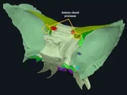
 3D representation of the sphenoid bone, with anterior clinoid processes circled | |
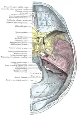
 Upper surface of the base of the skull (label for anterior clinoid process visible at center left. Sphenoid bone is yellow.) | |
| Details | |
| Identifiers | |
| Latin | Processus clinoideus anterior |
| TA98 | A02.1.05.022 |
| TA2 | 607 |
| FMA | 54693 |
| Anatomical terms of bone | |
The anterior clinoid process is a posterior projection of the sphenoid bone at the junction of the medial end of either lesser wing of sphenoid bone with the body of sphenoid bone. The bilateral processes flank the sella turcica anteriorly.
The ACP is an important structure for cranial and endovascular surgical operations for several structures, including the pituitary gland and the internal carotid artery.
Anatomy
The anterior clinoid process is a pyramid-shaped bony projection of the lesser wing of the sphenoid bone and forms part of the lateral wall of the optic canal. Between each ACP lies the sella turcica, which holds the pituitary gland. Additionally, the ACP is part of the anterior roof of the cavernous sinus. The posterior and inferior portions of the ACP border the internal carotid artery.[1][2]
Attachments
The free border of the tentorium cerebelli extends anteriorly on either side beyond the attached border of the same side (which ends anteriorly at the posterior clinoid process) to ultimately end by attaching onto the anterior clinoid process.[1][3]
Pathology
The anterior clinoid process projects over the internal carotid artery, which supplies the majority of blood to the brain. Because of the "spear-like" shape of the ACP and the large size of this artery, it is possible (though rare) that as a complication of major head trauma, the ACP may puncture the vessel and cause intracranial hemorrhage.[4] Additionally, when an aneurysm forms below the ACP, the projection may create a flat or pointed area of compression that may rupture the vessel.[5]
More frequently, this positioning means that the ACP is an important surgical access point for correction of aneurysms, tumors, and other operations in the structures adjacent to it. It is particularly common for surgery to involve the ACP when operating on giant pituitary adenomas[2] and giant meningiomas, a type of meningioma which may originate at the ACP or spread to it from other structures.[6]
Endovascular surgery has become an increasingly common procedure for lesions and aneurysms around the ACP, as the complex anatomy makes it difficult and dangerous to access via other surgical methods. However, endovascular surgery in the clinoid region is also relatively high-risk, so only serious injuries and diseases are indicated for this procedure.[2][7]
Etymology
The anterior and posterior clinoid processes surround the sella turcica like the four corners of a four poster bed. Cline is Greek for bed. –oid, as usual, indicates a similarity to.[8] The term may also come from the Greek root klinein or the Latin clinare, both meaning "sloped" as in "inclined".
Additional images
 Anterior clinoid process
References
![]() This article incorporates text in the public domain from page 151 of the 20th edition of Gray's Anatomy (1918)
This article incorporates text in the public domain from page 151 of the 20th edition of Gray's Anatomy (1918)
- 1 2 Waxman, S.G. (2024). "Chapter 11: Ventricles and Coverings of the Brain". Clinical Neuroanatomy (30th ed.). New York: McGraw Hill.
- 1 2 3 da Costa, Marcos Devanir Silva; de Oliveira Santos, Bruno Fernandes; de Araujo Paz, Daniel; Rodrigues, Thiago Pereira; Abdala, Nitamar; Centeno, Ricardo Silva; Cavalheiro, Sergio; Lawton, Michael T.; Chaddad-Neto, Feres (September 2016). "Anatomical Variations of the Anterior Clinoid Process: A Study of 597 Skull Base Computerized Tomography Scans". Operative Neurosurgery. 12 (3): 289–297. doi:10.1227/NEU.0000000000001138. ISSN 2332-4252.
- ↑ Sinnatamby, Chummy S. (2011). Last's Anatomy (12th ed.). p. 440. ISBN 978-0-7295-3752-0.
- ↑ Cheong, Jin Hwan; Kim, Jae Min; Kim, Choong Hyun (2011). "Bony Protuberances on the Anterior and Posterior Clinoid Processes Lead to Traumatic Internal Carotid Artery Aneurysm Following Craniofacial Injury". Journal of Korean Neurosurgical Society. 49 (1): 49. doi:10.3340/jkns.2011.49.1.49. ISSN 2005-3711. PMC 3070895. PMID 21494363.
- ↑ Nishizaki, Takafumi; Ikeda, Norio; Kurokawa, Yasushi; Okamura, Tomomi; Abiko, Seisho (October 2005). "Ruptured Internal Carotid Artery Anterior Wall Aneurysm Identified During Vasospasm: Case Report". Neurosurgery. 57 (4): E811. doi:10.1227/01.neu.0000178238.36637.43. ISSN 0148-396X.
- ↑ Attia, Moshe; Umansky, Felix; Paldor, Iddo; Dotan, Shlomo; Shoshan, Yigal; Spektor, Sergey (October 2012). "Giant anterior clinoidal meningiomas: surgical technique and outcomes". Journal of Neurosurgery. 117 (4): 654–665. doi:10.3171/2012.7.jns111675. ISSN 0022-3085.
- ↑ White, A; Roark, C; Case, D; Folzenlogen, Z; Kumpe, D; Ding, D; Seinfeld, J (August 2021). "E-001 Endovascular management of traumatic intracranial aneurysms from closed head injury". Journal of Neurointerventional Surgery. BMJ Publishing Group Ltd.: A62.1–A62. doi:10.1136/neurintsurg-2021-SNIS.97.
- ↑ "Anterior clinoid process of the sphenoid bone". AnatomyExpert. Retrieved 20 March 2013.
External links
- "Anatomy diagram: 34257.000-2". Roche Lexicon - illustrated navigator. Elsevier. Archived from the original on 2013-06-22.
- Anatomy photo:22:os-0910 at the SUNY Downstate Medical Center