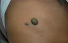
| Bleb (medicine) | |
|---|---|
 | |
| Blue rubber bleb nevus syndrome |
In medicine, a bleb is a blister-like protrusion (often hemispherical) filled with serous fluid. Blebs can form in a number of tissues by different pathologies, including frostbite and can "appear and disappear within a short time interval".
In pathology pulmonary blebs are small subpleural thin-walled air-containing spaces, not larger than 1-2 cm in diameter. Their walls are less than 1 mm thick. If they rupture, they allow air to escape into pleural space, resulting in a spontaneous pneumothorax.[1][2]
In ophthalmology, blebs may be formed intentionally in the treatment of glaucoma. In such treatments, functional blebs facilitate the circulation of aqueous humor, the blockage of which will lead to increase in eye pressure. Use of collagen matrix wound modulation device such as ologen during glaucoma surgery is known to produce vascular and functional blebs, which are positively correlated with treatment success rate.[3][4][5][6]
In the lungs, a bleb is a collection of air within the layers of the visceral pleura.
In breasts, a bleb is a milk blister (also known as blocked nipple pore, nipple blister, or “milk under the skin”).[7]
Pulmonary bleb
A pulmonary bleb is a small air collection found in the upper lobe of the lung located between the lung and the visceral pleura. When a bleb ruptures, air escapes into the chest cavity, resulting in a pneumothorax and possibly a collapsed lung. Bulla occurs when blebs grow larger or join together to create a larger cyst. There are usually no symptoms unless a pneumothorax occurs or the bulla grows very large. Blebs are usually associated with emphysema.[8]
External links
- Medical definition of bleb on MedicineNet.com
- Moorfields Bleb Grading System
References
- ↑ Ponuwei, Godwin A.; Dash, Phil R. (2016-12-01). "Bleb Formation in Human Fibrosarcoma HT1080 Cancer Cell Line Is Positively Regulated by the Lipid Signalling Phospholipase D2 (PLD2)". Achievements in the Life Sciences. 10 (2): 125–135. doi:10.1016/j.als.2016.11.001. ISSN 2078-1520.
- ↑ Charras, Guillaume; Paluch, Ewa (July 16, 2008). "Blebs lead the way: how to migrate without lamellipodia". Nature Reviews Molecular Cell Biology. Springer Science and Business Media LLC. 9 (9): 730–736. doi:10.1038/nrm2453. ISSN 1471-0072.
- ↑ Yuan F, Li L, Chen X, Yan X, Wang L (2015). "Biodegradable 3D-Porous Collagen Matrix (Ologen) Compared with Mitomycin C for Treatment of Primary Open-Angle Glaucoma: Results at 5 Years". J Ophthalmol. 2015: 637537. doi:10.1155/2015/637537. PMC 4452460. PMID 26078875.
{{cite journal}}: CS1 maint: multiple names: authors list (link) - ↑ Min JK, Kee CW, Sohn SW, Lee HJ, Woo JM, Yim JH (2013). "Surgical outcome of mitomycin C-soaked collagen matrix implant in trabeculectomy". J Glaucoma. 22 (6): 456–62. doi:10.1097/ijg.0b013e31826ab6b1. PMID 23263152. S2CID 20615016.
{{cite journal}}: CS1 maint: multiple names: authors list (link) - ↑ Boey PY, Narayanaswamy A, Zheng C, Perera SA, Htoon HM, Tun TA, Seah SK, Wong TT, Aung T (2011). "Imaging of blebs after phacotrabeculectomy with Ologen collagen matrix implants". Br J Ophthalmol. 95 (3): 340–4. doi:10.1136/bjo.2009.177758. PMID 20693559. S2CID 15912536.
{{cite journal}}: CS1 maint: multiple names: authors list (link) - ↑ Aptel F, Dumas S, Denis P (2009). "Ultrasound biomicroscopy and optical coherence tomography imaging of filtering blebs after deep sclerectomy with new collagen implant". Eur J Ophthalmol. 19 (2): 223–30. doi:10.1177/112067210901900208. PMID 19253238. S2CID 22594085.
{{cite journal}}: CS1 maint: multiple names: authors list (link) - ↑ White spot on the nipple
- ↑ Gaillard, Frank (February 16, 2021). "Radiology Reference Article". Radiopaedia. Retrieved November 18, 2023.