| Euglena gracilis | |
|---|---|
 | |
| Scientific classification | |
| Domain: | Eukaryota |
| Phylum: | Euglenozoa |
| Class: | Euglenoidea |
| Order: | Euglenales |
| Family: | Euglenaceae |
| Genus: | Euglena |
| Species: | E. gracilis |
| Binomial name | |
| Euglena gracilis Klebs, 1883 | |
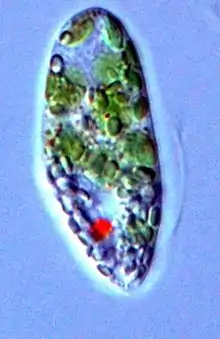
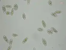
Euglena gracilis is a freshwater species of single-celled alga in the genus Euglena. It has secondary chloroplasts, and is a mixotroph able to feed by photosynthesis or phagocytosis. It has a highly flexible cell surface, allowing it to change shape from a thin cell up to 100 µm long to a sphere of approximately 20 µm. Each cell has two flagella, only one of which emerges from the flagellar pocket (reservoir) in the anterior of the cell, and can move by swimming, or by so-called "euglenoid" movement across surfaces. E. gracilis has been used extensively in the laboratory as a model organism, particularly for studying cell biology and biochemistry.[1]
Other areas of their use include studies of photosynthesis, photoreception, and the relationship of molecular structure to the biological function of subcellular particles, among others.[2] Euglena gracilis is the most studied member of the Euglenaceae.
Taxonomy

A morphological and molecular study of the Euglenozoa put E. gracilis in close kinship with the species Khawkinea quartana, with Peranema trichophorum basal to both,[3] although a later molecular analysis showed that E. gracilis was more closely related to Astasia longa than to certain other species recognized as Euglena.
The transcriptome of E. gracilis was sequenced, showing that E. gracilis has many unclassified genes which can make complex carbohydrates and natural products.[4][5]
Morphology
The morphology is characterized by a spindle-shaped cell with a length ranging from 40 to 150 micrometers. The cell contains a pellicle which is a flexible outer covering made up of proteinaceous strips called pellicular strips. This pellicle provides shape and structure to the cell. The movement of the E. gracilis is primarily achieved by its flagellum that emerges from a flagellar pocket. It has foward and backwards movement, as well as changes in its direction. Additionally, Euglena gracilis contains a light-sensitive eyespot, or stigma, which enables it to exhibit phototaxis by moving towards light sources for photosynthesis. The cell also possesses a contractile vacuole responsible for osmoregulation, helping maintain proper water balance within the cell. [6]
Energy storage
Paramylon is a unique storage polysaccharide found in Euglena gracilis, serving as a reserve carbohydrate for energy storage. Structurally, paramylon is a linear β-1,3-glucan, distinct from the storage polysaccharide starch of plants and some species of alga.[7]
The origin of the middle plastid membrane
The plastids contain three membranes. These membranes are an evolutionary vestige of the secondary endosymbiotic event that occurred between a phagotrophic eukaryovorous euglenid and a Pyramimonas-related green alga.[8] The plastids of Euglena are unusual since most secondary plastids are surrounded by four envelopes. The two inner ones are derived from the inner and outer chloroplast envelopes of the primary plastid of the alga that was taken up during the symbiotic event. The two outermost are derived from the plasma membrane of the alga (third) and the phagosome of the host (fourth).[8]
Biofuels
Microalgae are considered a possible source for biodiesel production due to their high lipid content. Its lipids may be suitable for biodiesel production due to their saturation, such as fatty acyl-CoA reductase and wax synthase. These ratios vary on environmental and cultivation conditions.[9]
References
- ↑ Russell, A. G.; Watanabe, Y; Charette, JM; Gray, MW (2005). "Unusual features of fibrillarin cDNA and gene structure in Euglena gracilis: Evolutionary conservation of core proteins and structural predictions for methylation-guide box C/D snoRNPs throughout the domain Eucarya". Nucleic Acids Research. 33 (9): 2781–91. doi:10.1093/nar/gki574. PMC 1126904. PMID 15894796.
- ↑ Wacker, Warren E. C. (1962-09-29). "Euglena: An Experimental Organism for Biochemical and Biophysical Studies". JAMA: The Journal of the American Medical Association. 181 (13): 1150. doi:10.1001/jama.1962.03050390052015. ISSN 0098-7484.
- ↑ Montegut-Felkner, Ann E.; Triemer, Richard E. (1997). "Phylogenetic Relationships of Selected Euglenoid Genera Based on Morphological and Molecular Data". Journal of Phycology. 33 (3): 512–9. Bibcode:1997JPcgy..33..512M. doi:10.1111/j.0022-3646.1997.00512.x. S2CID 83579360.
- ↑ "The potential in your pond". ScienceDaily. August 14, 2015. Retrieved December 14, 2023.
- ↑ O'Neill, Ellis C.; Trick, Martin; Hill, Lionel; Rejzek, Martin; Dusi, Renata G.; Hamilton, Christopher J.; Zimba, Paul V.; Henrissat, Bernard; Field, Robert A. (2015). "The transcriptome of Euglena gracilis reveals unexpected metabolic capabilities for carbohydrate and natural product biochemistry". Molecular BioSystems. 11 (10): 2808–21. doi:10.1039/C5MB00319A. PMID 26289754.
- ↑ Barsanti, Laura; Gualtieri, Paolo (2020-01-01), Konur, Ozcan (ed.), "Chapter 4 - Anatomy of Euglena gracilis", Handbook of Algal Science, Technology and Medicine, Academic Press, pp. 61–70, ISBN 978-0-12-818305-2, retrieved 2023-12-15
- ↑ Gissibl, Alexander; Sun, Angela; Care, Andrew; Nevalainen, Helena; Sunna, Anwar (2019). "Bioproducts From Euglena gracilis: Synthesis and Applications". Frontiers in Bioengineering and Biotechnology. 7. doi:10.3389/fbioe.2019.00108. ISSN 2296-4185. PMC 6530250. PMID 31157220.
- 1 2 Minorsky, Peter (2020-12-10). "On the Inside: The Origins of Euglena gracilis's Middle Plastid Envelope Membrane". Plantae. Retrieved 2023-12-15.
- ↑ Gissibl, Alexander; Sun, Angela; Care, Andrew; Nevalainen, Helena; Sunna, Anwar (2019). "Bioproducts From Euglena gracilis: Synthesis and Applications". Frontiers in Bioengineering and Biotechnology. 7. doi:10.3389/fbioe.2019.00108. ISSN 2296-4185. PMC 6530250. PMID 31157220.
External links
 Media related to Euglena gracilis at Wikimedia Commons