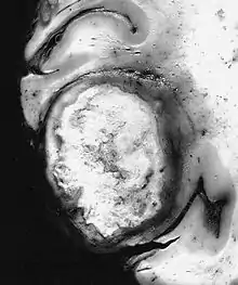
| Giant-cell glioblastoma | |
|---|---|
 Some glioblastomas, such as this giant-cell variant, are discrete firm masses which clinically and radiographically simulate metastatic carcinoma | |
| WHO Classification | |
| Standard name | Giant-cell glioblastoma |
| Structure | Neuroepithelial tumors └►Astrocytic tumors └►Glioblastoma └►Giant-cell glioblastoma |
| ICD-O Code & WHO Grade | |
| ICD-O Code | 9441/3 |
| WHO Grade | IV |
| Synonyms & Acronyms | |
| Synonyms | Monstrocellular sarcoma |
| Epidemiology | |
| Incidence | 0.15/100,000/y new cases/population/year |
| Age peak | 42 |
| M/F ratio | 1.6 |
| Prognosis | |
| Mean overall survival | 12 months |
| Medicine WikiProject/Neurology task force | |
The giant-cell glioblastoma is a histological variant of glioblastoma, presenting a prevalence of bizarre, multinucleated (more than 20 nuclei) giant (up to 400 μm diameter) cells.
It occasionally shows an abundant stromal reticulin network and presents a high frequency of TP53 gene mutations.[1][2]
Symptoms and signs are similar to those of the ordinary glioblastoma. Methodology of diagnosis and treatment are the same.
Prognosis is similar to the ordinary glioblastoma (about 12 months).[3] Some authors (see later) refer cases with a slightly better outcome.
Historical annotation
The giant-cell glioblastoma was originally termed "monstrocellular sarcoma", because of its stromal reticulin network,[4][5] but the astrocytic nature of the tumor was firmly established through the consistent GFAP expression analysis.[6][7][8]
Epidemiology
Incidence
The giant-cell glioblastoma is a rare neoplasia: its incidence is less than 1% of all brain tumors. It represents up to 5% of glioblastomas.[9]
Age and sex distribution
The mean age at clinical presentation is 42. The age distribution includes children and has a wider range than other diffuse astrocytomas (diffuse WHO grade II astrocytoma, anaplastic astrocytoma, ordinary glioblastoma).[9][10][11]
The giant-cell glioblastoma affects males more frequently. (The M/F ratio is 1.6.)[1]
Prognosis
Most patients with giant-cell glioblastoma have unfavourable prognosis,[12] but some authors report clinical results slightly better than the ordinary glioblastoma,[13][14][15][16][17] in all probability because this variant seems less infiltrative, due to the nature of giant cells of this type.[1]
See also
References
- 1 2 3 Ohgaki H, Peraud A, Nakazato Y, Watanabe K, von Deimling A (2000). "Giant cell glioblastoma". In Kleihues P, Cavenee WK (eds.). Pathology and Genetics of Tumours of the Nervous System. Lyon: IARC. ISBN 92-832-2409-4.
- ↑ Macchi G. (2005) [1981]. Malattie del sistema nervoso. PICCIN Editore. ISBN 88-299-1739-7.
- ↑ DeAngelis LM, Loeffler JS, Adam N, Mamelak AN (2007). "Primary and Metastatic Brain Tumors". In Pazdur R, Coia LR, Hoskins WJ, Wagman LD (eds.). Cancer Management: A Multidisciplinary Approach (10th ed.). Retrieved 4 August 2009.
- ↑ Zulch KJ (1979). Histological Typing of Tumours of the Central Nervous System. Geneva: World Health Organization. ISBN 978-92-4-176021-8. OCLC 6845931.
- ↑ Zulch KJ (1986). Brain Tumors: Their Biology and Pathology (3rd ed.). Berlin Heidelberg: Springer Verlag. ISBN 978-0-387-10933-6.
- ↑ Jacque CM, Kujas M, Poreau A, et al. (March 1979). "GFA and S 100 protein levels as an index for malignancy in human gliomas and neurinomas". Journal of the National Cancer Institute. 62 (3): 479–83. doi:10.1093/jnci/62.3.479. ISSN 0027-8874. PMID 216839.
- ↑ Kleihues P, Burger PC, Scheithauer BW (1993). Histological Typing of Tumours of the Central Nervous System (2nd ed.). Berlin Heidelberg: Springer Verlag. ISBN 978-3-540-56971-8.
- ↑ Russell DS, Rubinstein LJ (1989). Pathology of Tumors of the Nervous System (5th ed.). London: Edward Arnold.
- 1 2 Palma L, Celli P, Maleci A, Di Lorenzo N, Cantore G (1989). "Malignant monstrocellular brain tumours. A study of 42 surgically treated cases". Acta Neurochirurgica. 97 (1–2): 17–25. doi:10.1007/BF01577735. ISSN 0001-6268. PMID 2718792.
- ↑ Meyer-Puttlitz B, Hayashi Y, Waha A, et al. (September 1997). "Molecular genetic analysis of giant-cell glioblastomas". The American Journal of Pathology. 151 (3): 853–7. ISSN 0002-9440. PMC 1857850. PMID 9284834.
- ↑ Peraud A, Watanabe K, Plate KH, Yonekawa Y, Kleihues P, Ohgaki H (November 1997). "p53 mutations versus EGF receptor expression in giant-cell glioblastomas". Journal of Neuropathology and Experimental Neurology. 56 (11): 1236–41. doi:10.1097/00005072-199711000-00008. ISSN 0022-3069. PMID 9370234.
- ↑ Huang MC, Kubo O, Tajika Y, Takakura K (April 1996). "A clinico-immunohistochemical study of giant cell glioblastoma" (Free full text). Nōshuyō Byōri. 13 (1): 11–6. ISSN 0914-8108. PMID 8916121.
- ↑ Margetts JC, Kalyan-Raman UP (February 1989). "Giant-celled glioblastoma of brain. A clinico-pathological and radiological study of ten cases (including immunohistochemistry and ultrastructure)" (Free full text). Cancer. 63 (3): 524–31. doi:10.1002/1097-0142(19890201)63:3<524::AID-CNCR2820630321>3.0.CO;2-D. ISSN 0008-543X. PMID 2912529.
- ↑ Shinojima N, Kochi M, Hamada J, et al. (August 2004). "The influence of sex and the presence of giant cells on postoperative long-term survival in adult patients with supratentorial glioblastoma multiforme". Journal of Neurosurgery. 101 (2): 219–26. doi:10.3171/jns.2004.101.2.0219. ISSN 0022-3085. PMID 15309911.
- ↑ Becker DP, Benyo R, Roessmann U (January 1967). "Glial origin of monstrocellular tumor. Case report of prolonged survival" (Free full text). Journal of Neurosurgery. 26 (1): 72–7. doi:10.3171/jns.1967.26.1part1.0072. ISSN 0022-3085. PMID 6018784.
- ↑ Burger PC, Vollmer RT (September 1980). "Histologic factors of prognostic significance in the glioblastoma multiforme" (Free full text). Cancer. 46 (5): 1179–86. doi:10.1002/1097-0142(19800901)46:5<1179::AID-CNCR2820460517>3.0.CO;2-0. ISSN 0008-543X. PMID 6260329.
- ↑ Klein R, Mölenkamp G, Sörensen N, Roggendorf W (June 1998). "Favorable outcome of giant cell glioblastoma in a child. Report of an 11-year survival period". Child's Nervous System. 14 (6): 288–91. doi:10.1007/s003810050228. ISSN 0256-7040. PMID 9694343.