| Haemadin | |||||||||
|---|---|---|---|---|---|---|---|---|---|
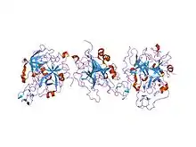
 crystal structure of the human alpha-thrombin-haemadin complex: an exosite ii-binding inhibitor | |||||||||
| Identifiers | |||||||||
| Symbol | Haemadin | ||||||||
| Pfam | PF09065 | ||||||||
| InterPro | IPR015150 | ||||||||
| SCOP2 | 1e0f / SCOPe / SUPFAM | ||||||||
| |||||||||
In molecular biology, haemadin is an anticoagulant peptide synthesised by the Indian leech, Haemadipsa sylvestris. It adopts a secondary structure consisting of five short beta-strands (beta1-beta5), which are arranged in two antiparallel distorted sheets formed by strands beta1-beta4-beta5 and beta2-beta3 facing each other. This beta-sandwich is stabilised by six enclosed cysteines arranged in a [1-2, 3-5, 4-6] disulfide pairing resulting in a disulfide-rich hydrophobic core that is largely inaccessible to bulk solvent. The close proximity of disulfide bonds [3-5] and [4-6] organises haemadin into four distinct loops. The N-terminal segment of this domain binds to the active site of thrombin, inhibiting it.[1]
Haemadin (MEROPS I14.002) belongs to a superfamily (MEROPS IM) of protease inhibitors that also includes hirudin (MEROPS I14.001) and antistasin (MEROPS I15).[2][3]
References
- ↑ Richardson JL, Kroger B, Hoeffken W, Sadler JE, Pereira P, Huber R, Bode W, Fuentes-Prior P (November 2000). "Crystal structure of the human alpha-thrombin-haemadin complex: an exosite II-binding inhibitor". EMBO J. 19 (21): 5650–60. doi:10.1093/emboj/19.21.5650. PMC 305786. PMID 11060016.
- ↑ "InterPro". www.ebi.ac.uk.
- ↑ "Clan IM". MEROPS - the Peptidase Database.