 | |
| Names | |
|---|---|
| Preferred IUPAC name
1-[6-(Dimethylamino)naphthalen-2-yl]dodecan-1-one | |
| Other names
Laurdan | |
| Identifiers | |
3D model (JSmol) |
|
| ChemSpider | |
PubChem CID |
|
| UNII | |
CompTox Dashboard (EPA) |
|
| |
| |
| Properties | |
| C24H35NO | |
| Molar mass | 353.55 |
| Appearance | Colourless solid |
| Melting point | 88 °C (190 °F; 361 K) (lit.) |
| DMF: soluble; acetonitrile: soluble, methanol: soluble | |
Except where otherwise noted, data are given for materials in their standard state (at 25 °C [77 °F], 100 kPa).
Infobox references | |
Laurdan is an organic compound which is used as a fluorescent dye when applied to fluorescence microscopy.[1][2] It is used to investigate membrane qualities of the phospholipid bilayers of cell membranes.[3][4][5][6] One of its most important characteristics is its sensitivity to membrane phase transitions as well as other alterations to membrane fluidity such as the penetration of water.[7][8]
History
Laurdan was first synthesized in 1979 by the Argentinian scientist Gregorio Weber, who started biomolecular fluorescence spectroscopy.[9] His thesis, "Fluorescence of Riboflavin, Diasphorase and Related Substances", was the starting point for the application of fluorescence spectroscopy to biomolecules.[9]
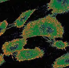
Laurdan was designed as a substitute for other dyes, such as previously modified lipids[10] that were inadequate to observe the membrane lipid bilayer because of their interaction with other compounds within the membrane lipid bilayer. Laurdan was designed specifically to study dipolar relaxation on cell membranes. Laurdan shows this effect more evidently because of its polar characteristics.[11][12] Laurdan was first applied to study membrane fluidity of live cells with a 2-Photon fluorescence microscope in 1994 [13] and it was found that the plasma membrane of cells is more rigid than that of the nuclear membrane.[13]
Chemical and physical properties
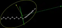
Laurdan is composed of a chain of lauric fatty acid (hydrophobic) linked to a naphthalene molecule.[14] Because of a partial charge separation between the 2-dimethylamino and the 6-carbonyl residues, the naphthalene moiety has a dipole moment, which increases upon excitation and causes the reorientation of the surrounding solvent dipoles. This causes its fluorescence and explains its importance in electronic microscopy.

The solvent’s reorientation requires energy. This energy requirement decreases the energy state of the excited probe, which is reflected in a continuous red shift in the probe’s emission spectrum. When the probe is in an apolar solvent the shift emission is blue, and a red-shifted emission is observed in polar solvents.
Due to its structure and its fluorescence characteristics, Laurdan is very useful in studies about lipid bilayer dynamics, more particularly about cell's plasmatic membrane's dynamics. The hydrophobic tail of the fatty acid allows the solubilization of the dye in the lipid bilayer, while the naphthalene moiety of the molecule stays at the level of the glycerol backbones of the membrane’s phospholipids. This means that the fluorescent part of the molecule is located towards the aqueous environment, which makes the reorientation of the solvent dipoles by Laurdan’s emission possible.
When Laurdan is located in the cell membrane its emission maximum is centered at 440 nm in gel-phase, and at 490 nm in liquid-phase. This spectral shift is the result of the dipolar relaxation of Laurdan on the lipidic environment, namely, the reorientation of solvents caused by Laurdan’s excitation. Particularly, due to some water molecules located at the level of the glycerol backbone, where the naphthalene moiety resides[15] which can only be reoriented in the liquid phase.
The geometry of the Laurdan molecule is as follows: the Dreiding energy, which is the energy related to the 3D structure of the molecule using the Dreiding force field,[16] is 71.47 kcal/mol. The volume is 377.73 Å3 while the minimal projection area is 53.09 Å2. The minimum z length is 24.09 Å, the maximal projection area is 126.21 Å2 and the maximum z length is 10.33 Å.[17]
Applications of Laurdan

Laurdan has the advantage of being able to be applied to living cells and therefore is able to provide information from complex membranes.
Due to its high sensitivity to the mobility and presence of solvent dipoles, changes in the emission spectrum can be calculated from the generalized polarization. Generalized polarization values vary from 1 (no solvent effect) to -1 (complete exposure to bulk water):[6] Laurdan anisotropy detects changes in plasma membrane fluidity caused by the interaction of determinate surroundings by calculating the generalized polarization and monitoring the reconstitution of lipid microdomains.[18][19]
The use of Laurdan as a fluorescent marker is to visualize and quantify the insolubility of the plasma membrane, analysing its remodelling activity. Rearrangements of glycosphingolipids, phospholipids, as well as cholesterol explains changes in membrane fluidity.[20]
Some studies developed at the Regional Center for Biotechnology at Haryana (India) have revealed that free hydroxyl groups on specific bile phospholipids increase solvent dipole penetration within the membrane. The number and order of these functional groups are tightly bound.[21]
Studies using mice have been of particular importance in sensing other biomolecules which influence glycerol and acyl chain regions of the plasma membrane. Dietary sources involved in the construction of lipid raft, n-3 PUFA from oil fish as well as polyphenols, affect the molecular and structural shape of the phospholipids in the membrane.[22][23] As such, this organisation model contributes to distinguishing effects of perturbations on cell membrane order and fluidity.[24]
See also
References
- ↑ Parasassi, T; Gratton E.; Levi M. (1997). "Two-photon fluorescence microscopy of laurdan generalized polarization domains in model and natural membranes". Biophys. J. 72 (6): 2413–2429. Bibcode:1997BpJ....72.2413P. doi:10.1016/S0006-3495(97)78887-8. PMC 1184441. PMID 9168019.
- ↑ Owen, D. M.; Neil. M. A.; Magee A. I. (2007). "Optical techniques for imaging membrane lipid microdomains in living cells". Seminars in Cell & Developmental Biology. 18 (5): 591–598. doi:10.1016/j.semcdb.2007.07.011. PMID 17728161.
- ↑ Bagatolli, L. A. (2006). "To see or not to see: lateral organization of biological membranes and fluorescence microscopy". Biochim. Biophys. Acta. 1758 (10): 1451–1456. doi:10.1016/j.bbamem.2006.05.019. PMID 16854370.
- ↑ Parasassi, T.; G. De Stasio; E. Gratton (1990). "Phase fluctuation in phospholipid membranes revealed by Laurdan fluorescence". Biophys. J. 57 (6): 1179–1186. Bibcode:1990BpJ....57.1179P. doi:10.1016/S0006-3495(90)82637-0. PMC 1280828. PMID 2393703.
- ↑ Pike, L. J. (2006). "Rafts defined: a report on the Keystone Symposium on Lipid Rafts and Cell Function". J. Lipid Res. 47 (7): 1597–1598. doi:10.1194/jlr.E600002-JLR200. PMID 16645198.
- 1 2 Sanchez, S. A.; M. A. Tricerri; E. Gratton (2007). "Laurdan generalized polarization: from cuvette to microscope". Modern Research and Educational Topics in Microscopy: 1007–1014.
- ↑ Gratton, S. A.; M. A. Tricerri, E. Gratton. (2012). "Laurdan generalized polarization fluctuations measures membrane packing micro-heterogeneity in vivo". Proc. Natl. Acad. Sci. USA. 109 (19): 7314–7319. Bibcode:2012PNAS..109.7314S. doi:10.1073/pnas.1118288109. PMC 3358851. PMID 22529342.
- ↑ Schneckenburger, P.; M. Wagner; H. Schneckenburger. (2010). "Fluorescence imaging of membrane dynamics in living cells". J. Biomed. 15 (4): 046017–046017–5. Bibcode:2010JBO....15d6017W. doi:10.1117/1.3470446. PMID 20799819.
- 1 2 Jameson, David M. (July 1998). "Gregorio Weber, 1916–1997: A Fluorescent Lifetime". Biophysical Journal. 75 (1): 419–421. Bibcode:1998BpJ....75..419J. doi:10.1016/s0006-3495(98)77528-9. PMC 1299713. PMID 9649401.
- ↑ Demchenko, Alexander P; Yves Mély; Guy Duportail; Andrey S. Klymchenko (6 May 2009). "Monitoring Biophysical Properties of Lipid Membranes by Environment- Sensitive Fluorescent Probes". Biophysical Journal. 96 (9): 3461–3470. Bibcode:2009BpJ....96.3461D. doi:10.1016/j.bpj.2009.02.012. PMC 2711402. PMID 19413953.
- ↑ Weber, G.; F. J. Farris (1979). "Synthesis and spectral properties of a hydrophobic fluorescent probe: 6 propionyl-2-(dimethylamino)naphthalene". Biochemistry. 18 (14): 3075–3078. doi:10.1021/bi00581a025. PMID 465454.
- ↑ Macgregor, R. B.; G.Weber. (1986). "Estimation of the polarity of the protein interior by optical spectroscopy". Nature. 319 (6048): 70–73. Bibcode:1986Natur.319...70M. doi:10.1038/319070a0. PMID 3941741. S2CID 4246323.
- 1 2 Yu, W; So, PT; French, T; Gratton, E (Feb 1996). "Fluorescence generalized polarization of cell membranes: a two-photon scanning microscopy approach". Biophys J. 70 (2): 626–36. Bibcode:1996BpJ....70..626Y. doi:10.1016/s0006-3495(96)79646-7. PMC 1224964. PMID 8789081.
- ↑ S. A. Sanchez, M.A.Tricerri, G. Gunther and E.Gratton, Lurdan GP:frm cuvette to mycroscope
- ↑ T. Parasassi, E.K. Krasnowska, L. Bagatolli, E. Gratton ,Journal of Fluorescence, 1998
- ↑ "Wiley online Library Static".
- ↑ "Chemicalize.org Laurdan Viewer".
- ↑ Ingelmo-Torres, M; Gaus, K.; Herms, A.; Gonzalez-Moreno, E.; Kassan, A. (2009). "Triton X-100 promotes cholesterol-dependent condensation of the plasma membrane" (PDF). Biochem. J. 420 (3): 373–381. doi:10.1042/BJ20090051. PMID 19309310.
- ↑ Harris, FM; Best, KB; Bell, JD (2002). "Use of laurdan fluorescence intensity and polarization to distinguish between changes in membrane fluidity and phospholipid order". Biochim. Biophys. Acta. 1565 (1): 123–8. doi:10.1016/s0005-2736(02)00514-x. PMID 12225860.
- ↑ Brignac-Huber, L.M; Reed, JR.; Eyer, MK.; Backes, WL. (2013). "Relationship between CYP1A2 Localization and Lipid Microdomain Formation as a Function of Lipid Composition". Drug Metab. Dispos. 41 (11): 1896–1905. doi:10.1124/dmd.113.053611. PMC 3807054. PMID 23963955.
- ↑ Streekanth, V; Bajaj, A.. (Oct 2013). "Number of free hydroxyl groups on bile Acid phospholipids determines the fluidity and hydration of model membranes". J. Phys. Chem. B. 117 (40): 12135–44. doi:10.1021/jp406340y. PMID 24079709.
- ↑ Kim, W; Barhoumi, R.; McMurray, DN.; Chapkin, RS. (2013). "Dietary fish oil and DHA down-regulate antigen-activated CD4+ T-cells while promoting the formation of liquid-ordered mesodomains". Br. J. Nutr. 111 (2): 254–60. doi:10.1017/S0007114513002444. PMC 4327854. PMID 23962659.
- ↑ Wesołowska, O; Gąsiorowska, J.; Petrus, J.; Czarnik-Matusewicz, B.; Michalak, K. (2013). "Interaction of prenylated chalcones and flavanones from common hop with phosphatidylcholine model membranes". Biochim. Biophys. Acta. 1838 (1 Pt B): 173–84. doi:10.1016/j.bbamem.2013.09.009. PMID 24060562.
- ↑ Di Venere, A; Nicolai, E.; Ivanov, I.; Dainese, E.; Adel, S.; Angelucci, BC.; Kuhn, H.; Maccarrone, M.; Mei, G. (2013). "Probing conformational changes in lipoxygenases upon membrane binding: Fine-tuning by the active site inhibitor ETYA". Biochim. Biophys. Acta. 1841 (1): 1–10. doi:10.1016/j.bbalip.2013.08.015. PMID 24012824.