| Medial pulvinar nucleus | |
|---|---|
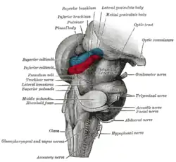
 Hind- and mid-brains; postero-lateral view. (Pulvinar visible near top.) | |
 Thalamic nuclei: MNG = Midline nuclear group AN = Anterior nuclear group MD = Medial dorsal nucleus VNG = Ventral nuclear group VA = Ventral anterior nucleus VL = Ventral lateral nucleus VPL = Ventral posterolateral nucleus VPM = Ventral posteromedial nucleus LNG = Lateral nuclear group PUL = Pulvinar MTh = Metathalamus LG = Lateral geniculate nucleus MG = Medial geniculate nucleus | |
| Details | |
| Part of | pulvinar |
| Identifiers | |
| Latin | nucleus pulvinaris medialis |
| Anatomical terms of neuroanatomy | |
Medial pulvinar nucleus (nucleus pulvinaris medialis) is one of four traditionally anatomically distinguished nuclei of the pulvinar of the thalamus. The other three nuclei of the pulvinar are called lateral, inferior and anterior pulvinar nuclei.
Connections
Afferent
- Medial pulvinar nucleus, together with its lateral and inferior nuclei, receives afferent input from superior colliculus.[1][2]
- Medial pulvinar nucleus also receives many afferent inputs from different cortical areas, including cingulate, posterior parietal, premotor and prefrontal cortical areas. This is the pattern of input connections typical for association relay nuclei of the thalamus.[3]
Efferent
- Medial pulvinar nucleus sends its widespread projections to the different areas of association cortex, including cingulate, posterior parietal, premotor and prefrontal cortical areas. This is the pattern of output connections typical for association relay nuclei of the thalamus.[3]
Functions
- Medial pulvinar nucleus, together with its lateral and inferior nuclei, is thought to be important for the initiation and compensation of saccadic movements of the eyes.[1][2] Those nuclei also participate in the visual attention regulation.[4][5]
- Medial pulvinar nucleus is also thought to participate in the process of integration and association of information received from different sensory modalities (multisensory or multimodal integration), and also in the process of integration and coordination of sensor input and its corresponding motor response (sensorimotor integration).[3]
Clinical significance
Lesions of the medial pulvinar nucleus can result in neglect syndromes and attentional deficits[6] while preserved connectivity to the medial pulvinar has been implicated in blindsight abilities.[7]
References
- 1 2 Berman R.; Wurtz R. (2011). "Signals conveyed in the pulvinar pathway from superior colliculus to cortical area mt". The Journal of Neuroscience. 31 (2): 373–384. doi:10.1523/jneurosci.4738-10.2011. PMC 6623455. PMID 21228149.
- 1 2 Robinson D.; Petersen S. (1985). "Responses of pulvinar neurons to real and self-induced stimulus movement". Brain Research. 338 (2): 392–394. doi:10.1016/0006-8993(85)90176-3. PMID 4027606. S2CID 7547426.
- 1 2 3 Cappe C.; Morel A.; Barone P.; Rouiller E.M. (2009). "The thalamocortical projection systems in primate: an anatomical support for multisensory and sensorimotor interplay". Cerebral Cortex. 19 (9): 2025–2037. doi:10.1093/cercor/bhn228. PMC 2722423. PMID 19150924.
- ↑ Petersen S.; Robinson D.; Morris J. (1987). "Contributions of the pulvinar to visual spatial attention". Neuropsychologia. 25 (1): 97–105. doi:10.1016/0028-3932(87)90046-7. PMID 3574654. S2CID 23143322.
- ↑ Chalupa, L. (1991). Visual function of the pulvinar. The Neural Basis of Visual Function. CRC Press, Boca Raton, Florida, pp. 140-159.
- ↑ Arend I.; Rafal R.; Ward R. (2008). "Spatial and temporal deficits are regionally dissociable in patients with pulvinar lesions". Brain. 131 (8): 2140–2152. doi:10.1093/brain/awn135. PMID 18669494.
- ↑ Kletenik I, Ferguson MA, Bateman JR, Cohen AL, Lin C, Tetreault A, Pelak VS, Anderson CA, Prasad S, Darby RR, Fox MD (2021-12-27). "Network Localization of Unconscious Visual Perception in Blindsight". Ann Neurol. 91 (2): 217–224. doi:10.1002/ana.26292. PMC 10013845. PMID 34961965. S2CID 245553461.
This article is issued from Wikipedia. The text is licensed under Creative Commons - Attribution - Sharealike. Additional terms may apply for the media files.