| Orthopneumovirus | |
|---|---|
 | |
| Pneumovirus structure and genome | |
 | |
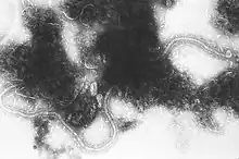
| Transmission electron micrograph of Human orthopneumovirus | |
| Virus classification | |
| (unranked): | Virus |
| Realm: | Riboviria |
| Kingdom: | Orthornavirae |
| Phylum: | Negarnaviricota |
| Class: | Monjiviricetes |
| Order: | Mononegavirales |
| Family: | Pneumoviridae |
| Genus: | Orthopneumovirus |
| Species | |
| |
The genus Orthopneumovirus consists of pathogens that target the upper respiratory tract within their specific hosts.[1] Every orthopneumovirus is characterized as host-specific, and has a range of diseases involved with respiratory illness. Orthopneumoviruses can cause diseases that range from a less-severe upper-respiratory illness to severe bronchiolitis or pneumonia. Orthopneumoviruses are found among sheep, cows, and most importantly humans. In humans, the orthopneumovirus that specifically impacts infants and small children is known as human respiratory syncytial virus.[2]
Taxonomy
| Genus | Species | Virus (Abbreviation) |
| Orthopneumovirus | Bovine orthopneumovirus | Bovine respiratory syncytial virus (BRSV) |
| Human orthopneumovirus | human respiratory syncytial virus A2 (HRSV-A2) | |
| human respiratory syncytial virus B1 (HRSV-B1) | ||
| Murine orthopneumovirus | murine pneumonia virus (MPV) | |
Other viruses in this taxon include canine pneumovirus.
Characteristics
The genus Orthopneumovirus is included in the family Pneumoviridae. Orthopneumoviruses are found specifically in the members of the species Homo sapiens, Ovis aries, Capra aegagrus hircus, Bos primigenius, and the order Rodentia.
The most common pneumoviruses are as follows:
•Homo sapiens: human respiratory syncytial virus
•Bos primigenius: bovine respiratory syncytial virus
•Rodentia: murine pneumonia virus
Symptoms
Mild symptoms may include rhinitis, coughing, and decreased appetite. More serious symptoms include wheezing, difficulty breathing, fever, bronchiolitis and pneumonia.[4]
Risk factors
Having a weak immune system and pre-existing conditions such as asthma can be leading factors in catching an orthopneumovirus. In elderly adults, having chronic heart or lung disease is also risk factor. Being in close proximity to a host who has been infected with an orthopneumovirus can also be a risk since most transmission happens via respiratory.
Diagnosis
Transmission of an infectious agent by another person or animal can be through blood, needles, blood transfusion, a mother to fetus, coughing, sneezing, saliva, or air transmission. Healthcare providers will determine the severity of the virus and possible treatment options. Healthcare providers will also decide if hospitalization is needed for more intense cases.[4][5][6]
Treatment
Treatment plans are not specific and are based upon a specific host's current symptoms. Pain relievers or nonsteroidal anti-inflammatory drugs such as acetaminophen or ibuprofen can be prescribed. Isolation is the first course of action for the infected host. In more serious cases hospitalization may be necessary and supplemental oxygen may be used to aid in oxygen intake. In severe viral detection, intubation and the use of a mechanical ventilation will be inserted as a breathing apparatus.[5]
Prevention
The best methods of prevention of orthopneumovirus infection are covering cough and sneezing to prevent transmission of possible pathogens. Isolating animals and humans that have a pneumovirus is the best way to prevent the virus from spreading. Molecular studies have been on the rise due to continuing materials and information obtained regarding recombinant DNA arrangements that might offer a foundation for vaccine development. Currently animals are able to receive vaccines specific to their virus strand. During cold and peak flu season, infants and small children have the option to receive monthly injections of small medication doses at a weak strength to help prevent virus/ host attachment.[7]
Cases
Human respiratory syncytial virus (HRSV) is the most known orthopneumovirus because of its direct correlation and importance in humans. RSV is the leading viral agent among pneumoviruses in pediatric upper respiratory diseases globally. New pneumoviruses have been discovered in the Netherlands among 28 children according to studies. Certain studies have isolated the children in hospitals to identify specific causes, contagion levels, and treatment options among those children.[2]
References
- 1 2 "ICTV Online (10th) Report".
- 1 2 Van Den Hoogen, Bernadette G.; De Jong, Jan C.; Groen, Jan; Kuiken, Thijs; De Groot, Ronald; Fouchier, Ron A.M.; Osterhaus, Albert D.M.E. (2001). "A newly discovered human pneumovirus isolated from young children with respiratory tract disease". Nature Medicine. 7 (6): 719–24. doi:10.1038/89098. PMC 7095854. PMID 11385510.
- ↑ Amarasinghe, Gaya K.; Bào, Yīmíng; Basler, Christopher F.; Bavari, Sina; Beer, Martin; Bejerman, Nicolás; Blasdell, Kim R.; Bochnowski, Alisa; Briese, Thomas (2017-04-07). "Taxonomy of the order Mononegavirales: update 2017". Archives of Virology. 162 (8): 2493–2504. doi:10.1007/s00705-017-3311-7. ISSN 1432-8798. PMC 5831667. PMID 28389807.
- 1 2 Bennett, Nicholas; Ellis, John; Bonville, Cynthia; Rosenberg, Helene; Domachowske, Joseph (2007). "Immunization strategies for the prevention of orthopneumovirus infections". Expert Review of Vaccines. 6 (2): 169–82. doi:10.1586/14760584.6.2.169. PMID 17408367. S2CID 44511976.
- 1 2 "Respiratory Syncytial Virus Infection (RSV)". Centers for Disease Control and Prevention. Retrieved 2019-06-03.
- ↑ McIntosh, K. M.; Chanock, R. M. (1985). "Respiratory Synoytial Virus". In Fields, Bernard N.; Knipe, David M.; Chanock, Robert M.; Hirsch, Martin S.; Melnick, Joseph L.; Monath, Thomas P.; Roizman, Bernard; Howley, Peter M.; Griffin, Diane E. (eds.). Fields Virology. New York: Raven Press. pp. 1285–304. ISBN 978-0-88167-552-8.
- ↑ Collins, Peter L. (1991). "The Molecular Biology of Human Respiratory Syncytial Virus (RSV) of the Genus Pneumovirus". In Kingsbury, David W. (ed.). The Paramyxoviruses. The Viruses. pp. 103–62. doi:10.1007/978-1-4615-3790-8_4. ISBN 978-1-4613-6689-8.