| Pectoralis minor | |
|---|---|
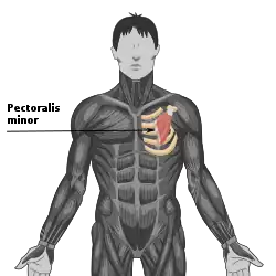
 Pectoralis minor (shown in red). | |
 Pectoralis minor muscle (shown in red). The bone shown in blue is the shoulder blade. | |
| Details | |
| Origin | Third to fifth ribs, near the costochondral junction |
| Insertion | Medial border and superior surface of the coracoid process of the scapula |
| Artery | Pectoral branch of the thoracoacromial trunk |
| Nerve | Medial pectoral nerve (C8) |
| Actions | Stabilizes the scapula by drawing it inferiorly and anteriorly against the thoracic wall, raises ribs in inhalation |
| Identifiers | |
| Latin | Musculus pectoralis minor |
| TA98 | A04.4.01.006 |
| TA2 | 2305 |
| FMA | 13109 |
| Anatomical terms of muscle | |
Pectoralis minor muscle (/ˌpɛktəˈrælɪs ˈmaɪnər/) is a thin, triangular muscle, situated at the upper part of the chest, beneath the pectoralis major in the human body. It arises from ribs III-V; it inserts onto the coracoid process of the scapula. It is innervated by the medial pectoral nerve. Its function is to stabilise the scapula by holding it fast in position against the chest wall.
Structure
Attachments
From the muscle's origin, the muscle's fibers pass superiorly and laterally, converging to form a flat tendon.
Origin
Pectoralis minor muscle arises from the upper margins and outer surfaces of the 3rd, 4th, and 5th ribs near their costal cartilages, and from the aponeuroses covering the intercostalis.[1]
Insertion
Its tendon inserts onto the medial border and upper surface of the coracoid process of the scapula.[1][2]
Innervation
The muscle receives motor innervation from the medial pectoral nerve.[3]
Relations
Pectoralis minor muscle forms part of the anterior wall of the axilla.[4] It is covered anteriorly (superficially) by the clavipectoral fascia. The medial pectoral nerve pierces the pectoralis minor and the clavipectoral fascia. In attaching to the coracoid process, the pectoralis minor forms a 'bridge' - structures passing into the upper limb from the thorax will pass directly underneath.[5]
Axillary nodes are classified according to their positions relative to the pectoralis minor muscle. Level 1 are lateral, Level 2 are deep, Level 3 are medial. The pectoralis minor divides the axillary artery into three parts (in contrary sequence compared to the nodes) - first part medial, second part deep/posterior, third part lateral in relation to the pectoralis minor.
Variations

The origin is from the second, third and fourth or fifth ribs. The tendon of insertion may extend over the coracoid process to the greater tubercle. It may be split into several parts. Absence of this muscle is rare but happens with certain uncommon diseases, such as the Poland syndrome.
Function
Pectoralis minor muscle depresses the point of the shoulder, drawing the scapula superior, towards the thorax, and throwing its inferior angle posteriorly.
See also
References
![]() This article incorporates text in the public domain from page 438 of the 20th edition of Gray's Anatomy (1918)
This article incorporates text in the public domain from page 438 of the 20th edition of Gray's Anatomy (1918)
- 1 2 Dommerholt, Jan (2011). "Chapter 34 - Dry needling of trigger points". Neck and Arm Pain Syndromes. Churchill Livingstone. pp. 430–438. doi:10.1016/B978-0-7020-3528-9.00034-0. ISBN 978-0-7020-3528-9.
- ↑ Bentley, J. Nicole; Yang, Linda J. S. (2015). Nerves and Nerve Injuries. Academic Press. pp. 563–574. doi:10.1016/B978-0-12-410390-0.00012-3. ISBN 978-0-12-410390-0.
- ↑ Maldonado, Kenia A.; Tadi, Prasanna (2022), "Anatomy, Thorax, Medial Pectoral Nerves", StatPearls, Treasure Island (FL): StatPearls Publishing, PMID 32310519, retrieved 2023-01-13
- ↑ Jacob, S. (2008-01-01). "Chapter 2 - Upper Limb". Human Anatomy. Churchill Livingstone. pp. 5–49. ISBN 978-0-443-10373-5.
- ↑ http://www.teachmeanatomy.com/muscles-of-the-pectoral-region/
External links
- Illustration: upper-body/pectoralis-minor from The Department of Radiology at the University of Washington
- Anatomy figure: 04:04-05 at Human Anatomy Online, SUNY Downstate Medical Center
- Anatomy figure: 05:02-08 at Human Anatomy Online, SUNY Downstate Medical Center
- Anatomy photo:05:ov-0200 at the SUNY Downstate Medical Center
- Anatomy photo:05:01-0102 at the SUNY Downstate Medical Center
- Slide