| Podoviridae | |
|---|---|
 | |
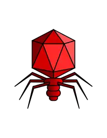
| Typical structure of a podovirus | |
| Virus classification | |
| (unranked): | Virus |
| Realm: | Duplodnaviria |
| Kingdom: | Heunggongvirae |
| Phylum: | Uroviricota |
| Class: | Caudoviricetes |
| Order: | Caudovirales |
| Family: | Podoviridae |
| Subfamilies and genera | |
|
see text | |
Podoviridae is a family of bacteriophage in the order Caudovirales often associated with T-7 like phages.[1] There are 130 species in this family, assigned to 3 subfamilies and 52 genera.[2][3] This family is characterized by having very short, noncontractile tails. Podoviradae are largely understudied and most new isolates are of the phicbkviruses genus, a group of giant viruses that appear to be Caulobacter specific.[1]
Structure


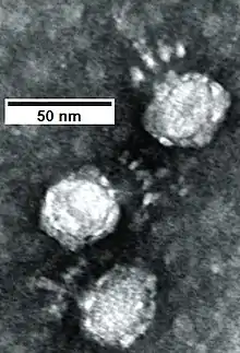
Viruses in Podoviridae are non-enveloped, with icosahedral and head-tail geometries. The diameter is around 60 nm,[2] and consists of 72 capsomers. The head protein has a molecular mass of ~38 kilodaltons and is present in 460 copies per virion. There are 9 structural proteins. The tail is non-contractile and has 6 short subterminal fibers. It is thick and rod-shaped and built of stacked disks. The maximum length is ~17 nm.
The double stranded DNA genome is linear, around 40-42kb in length,[2] and encodes ~55 genes. The guanine + cytosine content is ~50%. It has terminally redundant sequences and is nonpermuted. By weight, the genome constitutes ~50% of the viron. The genome encodes 9 structural proteins, an adenylated transferase B type DNA polymerase and an RNA polymerase. Three internal proteins constitute the polymerase complex. Two classes of genes are recognized (early and late). This classification is based on the timing of transcription that is temporally regulated. Genes with related functions are clustered together. Genome replication is bidirectional.
Life cycle
Viral replication is cytoplasmic. Entry into the host cell is achieved by adsorption into the host cell. Replication follows the DNA strand displacement model. DNA-templated transcription is the method of transcription. The virus exits the host cell by lysis, and holin/endolysin/spanin proteins. Bacteria serve as the natural host. Transmission routes are passive diffusion.[2] Phage infection is considered self-dosing thus self-limiting. After host lysis, new phages trigger a new infection cycle with surrounding bioavailable host species resulting in growth and expansion.[4]
Taxonomy
Genera within this family have ~40% identity between corresponding proteins. Subfamilies have ~20% identity between corresponding proteins.
The following subfamilies and their genera are recognized (-virinae denotes subfamily and -virus denotes genus):[3]
- Beephvirinae
- Flowerpowervirus
- Immanueltrevirus
- Manuelvirus
- Eekayvirinae
- Akonivirus
- Tinytimothyvirus
- Sepvirinae
- Diegovirus
- Oslovirus
- Traversvirus
The following genera are not assigned to a subfamily:[3]
- Anjalivirus
- Astrithrvirus
- Badaztecvirus
- Bjornvirus
- Bruynoghevirus
- Burrovirus
- Chopinvirus
- Cimandefvirus
- Delislevirus
- Dybvigvirus
- Enhodamvirus
- Enquatrovirus
- Fipvunavirus
- Firingavirus
- Gervaisevirus
- Giessenvirus
- Hollowayvirus
- Jasminevirus
- Kafunavirus
- Kelquatrovirus
- Kochitakasuvirus
- Kozyakovvirus
- Krylovvirus
- Kuravirus
- Lahexavirus
- Lastavirus
- Lederbergvirus
- Lessievirus
- Lightbulbvirus
- Myxoctovirus
- Pagevirus
- Parlovirus
- Perisivirus
- Privateervirus
- Rauchvirus
- Ryyoungvirus
- Schmidvirus
- Sendosyvirus
- Skarprettervirus
- Sortsnevirus
- Uetakevirus
- Vicosavirus
- Wumpquatrovirus
- Wumptrevirus
- Xuquatrovirus
Lastly, the species Pseudomonas virus 119X is unassigned to a genus or subfamily.[3]
References
- 1 2 Nguyen, Doreen; Ely, Bert (June 2018). "A Genome Comparison of T7-like Podoviruses That Infect Caulobacter crescentus". Current Microbiology. 75 (6): 760–765. doi:10.1007/s00284-018-1445-9. ISSN 1432-0991. PMID 29423729. S2CID 253813804.
- 1 2 3 4 "Viral Zone". ExPASy. Retrieved 1 July 2015.
- 1 2 3 4 "Virus Taxonomy: 2020 Release". International Committee on Taxonomy of Viruses (ICTV). March 2021. Retrieved 12 May 2021.
- ↑ Criscuolo, Elena; Spadini, Sara; Lamanna, Jacopo; Ferro, Mattia; Burioni, Roberto (2017). "Bacteriophages and Their Immunological Applications against Infectious Threats". Journal of Immunology Research. 2017: 3780697. doi:10.1155/2017/3780697. ISSN 2314-7156. PMC 5412166. PMID 28484722.