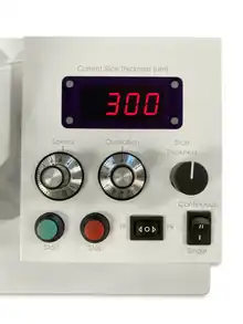
Precision-cut liver slices (PCLS) refer to thin sections of liver tissue obtained through a specialised cutting technique done with Compresstome vibratomes.[1] These slices are utilised to study drug metabolism, toxicity, and other aspects of liver function in a controlled laboratory setting.[2][3]

PCLS provide a robust ex vivo model; they maintain the liver's complex structure. PCLS are used for investigating mechanisms of liver injury and identifying therapeutic targets.[4][5][6]
History
Carlos Krumdieck introduced the Krumdieck tissue slicer for the first time in 1980.[7] This allowed rapid production of tissue slices. In 1985, PCLS were first introduced by Smith et al. as an in vitro model for toxicity testing. Initially the primary objectives of PCLS were metabolic studies and toxicity testing.[7] The focus of PCLS experiments shifted towards studies of chronic liver disease, such as fibrosis from 2005 onwards. The production of liver and intestinal tissue slices from humans and rats was described and published by de Graaf et al. in 2010. This technique is currently the standard procedure used by researchers for PCLS cultures.[7]
Basic preparation
Creating PCLS is a meticulous process that involves several essential steps. The use of Compresstome vibratomes is crucial in ensuring the production of precise and high-quality slices for research purposes.

Use of vibratomes

The basic steps involved in preparing PCLS using Compresstome vibratomes include:[8]
- Tissue selection
- Start by carefully selecting liver tissue from the desired species, such as rodents or humans, ensuring the tissue is of high quality and health.
- Tissue embedding
- To facilitate slicing and maintain tissue structure, the liver tissue is typically embedded in a suitable medium, such as agarose or gelatin, into the specimen holder of the Compresstome vibratome.
- Slicing process
- The vibratome operates by oscillating a blade vertically at high frequencies while the tissue is submerged in a cutting solution. This mechanical oscillation creates thin and precise slices of tissue. Researchers can adjust cutting parameters, such as slice thickness, to meet specific experimental requirements. Typically, PCLS have thicknesses ranging from 200–500µm.
- Post-processing
- Depending on the research objectives, PCLS may undergo additional steps such as washing, culturing, or treatment with substances of interest, such as drugs or stimuli.
Vibrating microtome
Compresstome vibratome provides high throughput PCLS. It allows the rapid and precise preparation of tissue slices, significantly reducing the time and effort required for sectioning large numbers of samples. This capability is particularly beneficial for laboratories and research institutions engaged in drug screening, toxicology studies, and extensive tissue-based experiments. The Compresstome is capable of sectioning at least 30 core samples of tissue simultaneously.[9]
Advantages of PCLS
There are several benefits of using PCLS in medication development and research. They offer more physiologically possible models compared to standard cell culture. More accurate evaluations of drug metabolism, toxicity, and overall liver function are made possible by this approach, which maintains tissue architecture, cell-cell connections, and metabolic processes.[10] PCLS help researchers to understand medication processes and potential side effects by allowing them to examine how drugs affect particular types of liver cells. Slice thickness uniformity improves accuracy and is a useful tool for in vitro research that closely resembles in vivo settings.[4]
Limitations of PCLS
PCLS have several limitations, such as only lasting a few hours.[11] This may limit the duration of the research and make it difficult to see the long-term effects of some drugs. Furthermore, slicing and preparing the slices may cause stress reactions, which might affect the results of the experiment. It might be difficult, but maintaining physiological oxygen levels and nutrition supplies during the experiment is essential. These drawbacks highlight the necessity of cautious experimental planning and interpretation when using PCLS in studies.[12]
References
- ↑ https://www.jhep-reports.eu/article/S2589-5559(22)00037-4/fulltext
- ↑ Palma, E.; Doornebal, E. J.; Chokshi, S. (2019). "Precision-cut liver slices: A versatile tool to advance liver research". Hepatology International. 13 (1): 51–57. doi:10.1007/s12072-018-9913-7. PMC 6513823. PMID 30515676.
- ↑ Van De Bovenkamp, M.; Groothuis, G.M.M.; Meijer, D.K.F.; Olinga, P. (2007). "Liver fibrosis in vitro: Cell culture models and precision-cut liver slices". Toxicology in Vitro. 21 (4): 545–557. doi:10.1016/j.tiv.2006.12.009. PMID 17289342.
- 1 2 Dewyse, Liza; De Smet, Vincent; Verhulst, Stefaan; Eysackers, Nathalie; Kunda, Rastislav; Messaoudi, Nouredin; Reynaert, Hendrik; Van Grunsven, Leo A. (2022). "Improved Precision-Cut Liver Slice Cultures for Testing Drug-Induced Liver Fibrosis". Frontiers in Medicine. 9. doi:10.3389/fmed.2022.862185. PMC 9007724. PMID 35433753.
- ↑ Lerche-Langrand, Carole; Toutain, Herve J. (2000). "Precision-cut liver slices: Characteristics and use for in vitro pharmaco-toxicology". Toxicology. 153 (1–3): 221–253. doi:10.1016/S0300-483X(00)00316-4. PMID 11090959.
- ↑ Brugger, Marcus; Laschinger, Melanie; Lampl, Sandra; Schneider, Annika; Manske, Katrin; Esfandyari, Dena; Hüser, Norbert; Hartmann, Daniel; Steiger, Katja; Engelhardt, Stefan; Wohlleber, Dirk; Knolle, Percy A. (2022). "High precision-cut liver slice model to study cell-autonomous antiviral defense of hepatocytes within their microenvironment". Jhep Reports. 4 (5). doi:10.1016/j.jhepr.2022.100465. PMC 9019249. PMID 35462860.
- 1 2 3 Dewyse, L.; Reynaert, H.; Van Grunsven, L. A. (2021). "Best Practices and Progress in Precision-Cut Liver Slice Cultures". International Journal of Molecular Sciences. 22 (13): 7137. doi:10.3390/ijms22137137. PMC 8267882. PMID 34281187.
- ↑ Dewyse, Liza; Reynaert, Hendrik; Van Grunsven, Leo A. (2021). "Best Practices and Progress in Precision-Cut Liver Slice Cultures". International Journal of Molecular Sciences. 22 (13): 7137. doi:10.3390/ijms22137137. PMC 8267882. PMID 34281187.
- ↑ Dewyse, L.; De Smet, V.; Verhulst, S.; Eysackers, N.; Kunda, R.; Messaoudi, N.; Reynaert, H.; Van Grunsven, L. A. (2022). "Improved Precision-Cut Liver Slice Cultures for Testing Drug-Induced Liver Fibrosis". Frontiers in Medicine. 9. doi:10.3389/fmed.2022.862185. PMC 9007724. PMID 35433753.
- ↑ Ramm, Grant A.; Tirnitz-Parker, Janina E. E.; Olynyk, John K.; Gobert, Geoffrey N.; Nawaratna, Sujeevi K.; Fernandez-Rojo, Manuel A.; Gratte, Francis D.; Lim, Hong Kiat; Pearen, Michael A. (2020). "Murine Precision-Cut Liver Slices as an Ex Vivo Model of Liver Biology". Journal of Visualized Experiments (157): e60992. doi:10.3791/60992. PMID 32225165. S2CID 214734813.
- ↑ De Graaf, Inge A M.; Olinga, Peter; De Jager, Marina H.; Merema, Marjolijn T.; De Kanter, Ruben; Van De Kerkhof, Esther G.; Groothuis, Geny M M. (2010). "Preparation and incubation of precision-cut liver and intestinal slices for application in drug metabolism and toxicity studies". Nature Protocols. 5 (9): 1540–1551. doi:10.1038/nprot.2010.111. PMID 20725069. S2CID 12512305.
- ↑ Palma, Elena; Doornebal, Ewald Jan; Chokshi, Shilpa (2019). "Precision-cut liver slices: A versatile tool to advance liver research". Hepatology International. 13 (1): 51–57. doi:10.1007/s12072-018-9913-7. PMC 6513823. PMID 30515676.