Silicateins are enzymes which catalyse the formation of biosilica from monomeric silicon compounds (such as silicic acid) extracted from the natural environment.[1] Environmental silicates are absorbed by specific biota, including diatoms, radiolaria, silicoflagellates, and siliceous sponges; silicateins have so far only been found in sponges. Silicateins are homologous to the cysteine protease cathepsin.[2][3]
In sponges, the silicatein enzymes reside in the axial filaments of the axial canals of the siliceous spicules.[1]
In contrast, diatoms do not use silicateins but rather small specialised peptides called silaffins which attach long chain polyamines (LCPAs) to lysine groups. Free LCPAs can also cooperate with silaffins.[2] Both silicateins and silaffins form higher-order structures which act both as structural templates (for exoskeletons) and mechanistic catalysts for the polycondensation reactions of silicon-compounds.
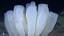
The Venus' flower basket siliceous sponge is a well-known example of an organism that utilises silicatein. It is known for its remarkable ability to extract silicic acid from surrounding seawater, which is then converted into complex 3D silica structures at ambient temperatures underwater, something human engineering capabilities are unable to replicate without the use of high-temperature.[4]
Another example of silicatein-utilising organisms are the suberites, a genus of sea sponge in the family Suberitidae.[5] Suberites consist mostly of cells, in contrast with other Porifera (such as the class Hexactinellida, to which the Venus' flower basket belongs) which are syncytial.[6] The extracellular matrix of siliceous spicules give suberites their structural foundation; these consist of bio-silica, a silicon dioxide polymer.[7] These inorganic structures provide support for the animals.[8][7][9][10] Silica deposition begins intracellularly and is carried out by the enzyme silicatein.[7][8][11] Silicateins are modulated by a group of proteins called silintaphins[12] The process occurs in specialized cells known as sclerocytes.[8]
Lubomirskia baikalensis, also known as Lake Baikal sponge, has been studied to explore the gene family of silicateins and their role in the morphogenesis of these sponges.[13]
References
- 1 2 Müller, W.E., Boreiko, A., Wang, X., Belikov, S.I., Wiens, M., Grebenjuk, V.A., Schloβmacher, U. and Schröder, H.C., 2007. Silicateins, the major biosilica forming enzymes present in demosponges: protein analysis and phylogenetic relationship. Gene, 395(1-2), pp.62-71. doi:10.1016/j.gene.2007.02.014
- 1 2 Otzen D. (2012). The role of proteins in biosilicification. Scientifica, 2012, 867562. doi:10.6064/2012/867562
- ↑ Shimizu, K., Cha, J., Stucky, G.D. and Morse, D.E., 1998. Silicatein α: cathepsin L-like protein in sponge biosilica. Proceedings of the National Academy of Sciences, 95(11), pp.6234-6238. doi:10.1073/pnas.95.11.6234
- ↑ Bullis, Kevin (1 November 2006). "Silicon and Sun". MIT Technology Review. Retrieved 6 May 2020.
- ↑ "Suberites Nardo, 1833". WoRMS - World Register of Marine Species. Retrieved 6 May 2020.
- ↑ W. Muller, Review: How was metazoan threshold crossed? The hypothetical Urmetazoa. Comparative Biochemistry and Physiology Part A 129, 433 (2001). doi:10.1016/s1095-6433(00)00360-3
- 1 2 3 W. Xiaohong et al., Evagination of Cells Controls Bio-Silica Formation and Maturation during Spicule Formation in Sponges. PLoS ONE 6, 1 (2011). doi:10.1371/journal.pone.0020523
- 1 2 3 X. Wang et al., Silicateins, silicatein interactors and cellular interplay in sponge skeletogenesis: formation of glass fiber-like spicules. FEBS Journal 279, 1721 (2012) https:/doi.org10.1111/j.1742-4658.2012.08533.x
- ↑ W. E. G. Müller et al., Hardening of bio-silica in sponge spicules involves an aging process after its enzymatic polycondensation: Evidence for an aquaporin-mediated water absorption. BBA − General Subjects 1810, 713 (2011). doi:10.1016/j.bbagen.2011.04.009
- ↑ W. E. G. Müller et al., Silicateins, the major biosilica forming enzymes present in demosponges: Protein analysis and phylogenetic relationship. Gene 395, 62 (2007). doi:10.1016/j.gene.2007.02.014
- ↑ W. E. G. Müller et al., Identification of a silicatein(-related) protease in the giant spicules of the deep-sea hexactinellid Monorhaphis chuni. Journal of Experimental Biology 211, 300 (2008) doi:10.1242/jeb.008193
- ↑ W. E. G. Müller et al., The silicatein propeptide acts as inhibitor/modulator of self-organization during spicule axial filament formation. FEBS Journal 280, 1693 (2013). doi:10.1111/febs.12183
- ↑ Belikov; Kaluzhnaya; Schröder; Müller; and Müller (2007). Lake Baikal endemic sponge Lubomirskia baikalensis: structure and organization of the gene family of silicatein and its role in morphogenesis. Porifera Research: Biodiversity, Innovation and Sustainability, pp. 179-188
![]() This article incorporates text by Otzen, Daniel available under the CC BY 3.0 license.
This article incorporates text by Otzen, Daniel available under the CC BY 3.0 license.