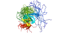
PR domain containing 16, also known as PRDM16, is a protein which in humans is encoded by the PRDM16 gene.[5][6]
PRDM16 acts as a transcription coregulator that controls the development of brown adipocytes in brown adipose tissue.[7] Previously, this coregulator was believed to be present only in brown adipose tissue, but more recent studies have shown that PRDM16 is highly expressed in subcutaneous white adipose tissue as well.[7]
Function
The protein encoded by this gene is a zinc finger transcription factor.[6] PRDM16 controls the cell fate between muscle and brown fat cells. Loss of PRDM16 from brown fat precursors causes a loss of brown fat characteristics and promotes muscle differentiation.[8]
Clinical significance
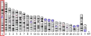
The reciprocal translocation t(1;3)(p36;q21) occurs in a subset of myelodysplastic syndrome (MDS) and acute myeloid leukemia (AML). This gene is located near the 1p36.3 breakpoint and has been shown to be specifically expressed in the t(1:3)(p36;q21)-positive MDS/AML. The protein encoded by this gene contains an N-terminal PR domain. The translocation results in the overexpression of a truncated version of this protein that lacks the PR domain, which may play an important role in the pathogenesis of MDS and AML. Alternatively spliced transcript variants encoding distinct isoforms have been reported.[6]
PRDM16 in BAT
Brown adipose tissue (BAT) oxidizes chemical energy to produce heat. This heat energy can act as a defense against hypothermia and obesity.[7] PRDM16 is highly enriched in brown adipose cells as compared to white adipose cells, and plays a role in these thermogenic processes in brown adipose tissue. PRDM16 activates brown fat cell identity and can control the determination of brown adipose fate. A knock-out of PRDM16 in mice shows a loss of brown cell characteristics, showing that PRDM16 activity is important in determining brown adipose fate.[9] Brown adipocytes consist of densely packed mitochondria that contain uncoupling protein 1 (UCP-1). UCP-1 plays a key role in brown adipocyte thermogenesis. The presence of PRDM16 in adipose tissue causes a significant up-regulation of thermogenic genes, such as UCP-1 and CIDEA, resulting in thermogenic heat production.[7] Understanding and stimulating the thermogenic processes in brown adipocytes provides possible therapeutic options for treating obesity.[9]
PRDM16 in WAT
White adipose tissue (WAT) primarily stores excess energy in the form of triglycerides.[7][9] Recent research has shown that PRDM16 is present in subcutaneous white adipose tissue.[7] The activity of PRDM16 in white adipose tissue leads to the production of brown fat-like adipocytes within white adipose tissue, called beige cells (also called brite cells). These beige cells have a brown adipose tissue-like phenotype and actions, including thermogenic processes seen in BAT.[7] In mice, the levels of PRDM16 within WAT, specifically anterior subcutaneous WAT and inguinal subcutaneous WAT, is about 50% that of interscapular BAT, both in protein expression and in mRNA quantity.[7] This expression takes place primarily within mature adipocytes. Transgenic aP2-PRDM16 mice were used in a study to observe the effects of PRDM16 expression in WAT.[7] The study found that the presence of PRDM16 in subcutaneous WAT leads to a significant up-regulation of brown-fat selective genes UCP-1, CIDEA, and PPARGC1A. This up-regulation lead to the development of a BAT-like phenotype within the white adipose tissue. Expression of PRDM16 has also been shown to protect against high-fat diet induced weight gain.[7] Seale et al.’s experiment with aP2-PRDM16 transgenic mice and wild type mice showed that transgenic mice eating a 60% high-fat diet had significantly less weight gain than wild type mice on the same diet. Seale et al. determined the weight difference was not due to differences in food intake, as both transgenic and wild type mice were consuming the same amount of food on a daily basis. Rather, the weight difference stemmed from higher energy expenditure in the transgenic mice. Another of Seale et al.’s experiments showed the transgenic mice consumed a greater volume of oxygen over a 72-hour period than the wild type mice, showing a greater amount of energy expenditure in the transgenic mice.[7] This energy expenditure in turn is attributed to PRDM16’s ability to up-regulate UCP-1 and CIDEA gene expression, resulting in thermogenesis.
If human WAT expresses PRDM16 as in mice, this WAT could be a potential target for stimulating energy expenditure and combating obesity.
Notes
- 1 2 3 GRCh38: Ensembl release 89: ENSG00000142611 - Ensembl, May 2017
- 1 2 3 GRCm38: Ensembl release 89: ENSMUSG00000039410 - Ensembl, May 2017
- ↑ "Human PubMed Reference:". National Center for Biotechnology Information, U.S. National Library of Medicine.
- ↑ "Mouse PubMed Reference:". National Center for Biotechnology Information, U.S. National Library of Medicine.
- ↑ Mochizuki N, Shimizu S, Nagasawa T, Tanaka H, Taniwaki M, Yokota J, Morishita K (November 2000). "A novel gene, MEL1, mapped to 1p36.3 is highly homologous to the MDS1/EVI1 gene and is transcriptionally activated in t(1;3)(p36;q21)-positive leukemia cells". Blood. 96 (9): 3209–14. doi:10.1182/blood.V96.9.3209. PMID 11050005.
- 1 2 3 "Entrez Gene: PRDM16 PR domain containing 16".
- 1 2 3 4 5 6 7 8 9 10 11 Patrick Seale, Heather M. Conroe, Jennifer Estall, Shingo Kajimura, Andrea Frontini, Jeff Ishibashi, Paul Cohen, Saverio Cinti, Bruce M. Spiegelman (January 2011). "Prdm16 determines the thermogenic program of subcutaneous white adipose tissue in mice". The Journal of Clinical Investigation. 121 (1): 96–105. doi:10.1172/JCI44271. PMC 3007155. PMID 21123942.
{{cite journal}}: CS1 maint: overridden setting (link) - ↑ Seale P, Bjork B, Yang W, Kajimura S, Chin S, Kuang S, Scimè A, Devarakonda S, Conroe HM, Erdjument-Bromage H, Tempst P, Rudnicki MA, Beier DR, Spiegelman BM (August 2008). "PRDM16 controls a brown fat/skeletal muscle switch". Nature. 454 (7207): 961–7. Bibcode:2008Natur.454..961S. doi:10.1038/nature07182. PMC 2583329. PMID 18719582.
- 1 2 3 Patrick Seale, Shingo Kajimura, Wenli Yang, Sherry Chin, Lindsay Rohas, Marc Uldry, Geneviève Tavernier, Dominique Langin, Bruce M Spiegelman (July 2007). "Transcriptional Control of Brown Fat Determination by PRDM16". Cell Metabolism. 6 (1): 38–54. doi:10.1016/j.cmet.2007.06.001. PMC 2564846. PMID 17618855.
{{cite journal}}: CS1 maint: overridden setting (link)
References
- This article incorporates text from the United States National Library of Medicine, which is in the public domain.
- Kajimura S (2009). "Initiation of myoblast to brown fat switch by a PRDM16–C/EBP-β transcriptional complex". Nature. 460 (7259): 1154–1158. Bibcode:2009Natur.460.1154K. doi:10.1038/nature08262. PMC 2754867. PMID 19641492.
Further reading
- Nakajima D, Okazaki N, Yamakawa H, et al. (2003). "Construction of expression-ready cDNA clones for KIAA genes: manual curation of 330 KIAA cDNA clones". DNA Res. 9 (3): 99–106. doi:10.1093/dnares/9.3.99. PMID 12168954.
- Bloomfield CD, Garson OM, Volin L, et al. (1986). "t(1;3)(p36;q21) in acute nonlymphocytic leukemia: a new cytogenetic-clinicopathologic association". Blood. 66 (6): 1409–13. doi:10.1182/blood.V66.6.1409.1409. PMID 4063527.
- Secker-Walker LM, Mehta A, Bain B (1996). "Abnormalities of 3q21 and 3q26 in myeloid malignancy: a United Kingdom Cancer Cytogenetic Group study". Br. J. Haematol. 91 (2): 490–501. doi:10.1111/j.1365-2141.1995.tb05329.x. PMID 8547101. S2CID 23922912.
- Mochizuki N, Shimizu S, Nagasawa T, et al. (2000). "A novel gene, MEL1, mapped to 1p36.3 is highly homologous to the MDS1/EVI1 gene and is transcriptionally activated in t(1;3)(p36;q21)-positive leukemia cells". Blood. 96 (9): 3209–14. doi:10.1182/blood.V96.9.3209. PMID 11050005.
- Nagase T, Kikuno R, Hattori A, et al. (2001). "Prediction of the coding sequences of unidentified human genes. XIX. The complete sequences of 100 new cDNA clones from brain which code for large proteins in vitro". DNA Res. 7 (6): 347–55. doi:10.1093/dnares/7.6.347. PMID 11214970.
- Strausberg RL, Feingold EA, Grouse LH, et al. (2003). "Generation and initial analysis of more than 15,000 full-length human and mouse cDNA sequences". Proc. Natl. Acad. Sci. U.S.A. 99 (26): 16899–903. Bibcode:2002PNAS...9916899M. doi:10.1073/pnas.242603899. PMC 139241. PMID 12477932.
- Xinh PT, Tri NK, Nagao H, et al. (2003). "Breakpoints at 1p36.3 in three MDS/AML(M4) patients with t(1;3)(p36;q21) occur in the first intron and in the 5' region of MEL1". Genes Chromosomes Cancer. 36 (3): 313–6. doi:10.1002/gcc.10176. PMID 12557231. S2CID 36946681.
- Nishikata I, Sasaki H, Iga M, et al. (2004). "A novel EVI1 gene family, MEL1, lacking a PR domain (MEL1S) is expressed mainly in t(1;3)(p36;q21)-positive AML and blocks G-CSF-induced myeloid differentiation". Blood. 102 (9): 3323–32. doi:10.1182/blood-2002-12-3944. PMID 12816872.
- Yoshida M, Nosaka K, Yasunaga J, et al. (2004). "Aberrant expression of the MEL1S gene identified in association with hypomethylation in adult T-cell leukemia cells". Blood. 103 (7): 2753–60. doi:10.1182/blood-2003-07-2482. hdl:2433/147510. PMID 14656887.
- Ota T, Suzuki Y, Nishikawa T, et al. (2004). "Complete sequencing and characterization of 21,243 full-length human cDNAs". Nat. Genet. 36 (1): 40–5. doi:10.1038/ng1285. PMID 14702039.
- Lahortiga I, Agirre X, Belloni E, et al. (2004). "Molecular characterization of a t(1;3)(p36;q21) in a patient with MDS. MEL1 is widely expressed in normal tissues, including bone marrow, and it is not overexpressed in the t(1;3) cells". Oncogene. 23 (1): 311–6. doi:10.1038/sj.onc.1206923. hdl:10171/19578. PMID 14712237.
- Ott MG, Schmidt M, Schwarzwaelder K, et al. (2006). "Correction of X-linked chronic granulomatous disease by gene therapy, augmented by insertional activation of MDS1-EVI1, PRDM16 or SETBP1". Nat. Med. 12 (4): 401–9. doi:10.1038/nm1393. PMID 16582916. S2CID 7601162.
- Stevens-Kroef MJ, Schoenmakers EF, van Kraaij M, et al. (2006). "Identification of truncated RUNX1 and RUNX1-PRDM16 fusion transcripts in a case of t(1;21)(p36;q22)-positive therapy-related AML". Leukemia. 20 (6): 1187–9. doi:10.1038/sj.leu.2404210. PMID 16598304. S2CID 40770542.
- Stiffler MA, Grantcharova VP, Sevecka M, MacBeath G (2007). "Uncovering quantitative protein interaction networks for mouse PDZ domains using protein microarrays". J. Am. Chem. Soc. 128 (17): 5913–22. doi:10.1021/ja060943h. PMC 2533859. PMID 16637659.
External links
- PRDM16+protein,+human at the U.S. National Library of Medicine Medical Subject Headings (MeSH)


